Track neural activity, network maturation, and functional phenotype over time with noninvasive MEA recordings.
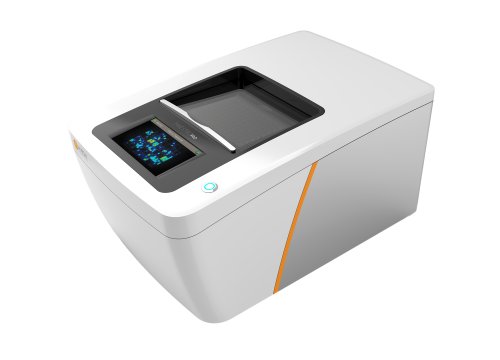
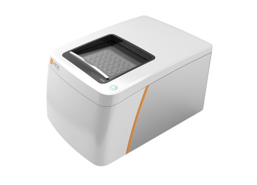
Human stem cell-derived neurons can express expected markers and morphology without necessarily developing mature electrical activity. Maestro MEA provides a functional readout of firing, bursting, synchrony, oscillations, and local field potentials, helping researchers characterize how neural models develop and respond to culture conditions.
-
Functionally optimize stem cell differentiation and culture conditions>
-
Track network maturation with spikes and local field potentials>
-
Compare functional phenotypes across neural models>
-
A longitudinal workflow for stem cell-derived neural models>
Stem cell-derived neural models can vary substantially depending on differentiation protocol, media, culture duration, cell density, and other experimental conditions.
MEA provides a quantitative way to determine how those variables affect functional maturation.
In the study shown on this page, hPSC-derived neurons were monitored for 18 weeks under different media conditions. Network bursts first appeared at week 6 in both BrainPhys and DMEM/F-12/NB-A cultures. By week 18, BrainPhys cultures produced 114 network bursts during a 10-minute recording compared with 54 in DMEM/F-12/NB-A cultures.
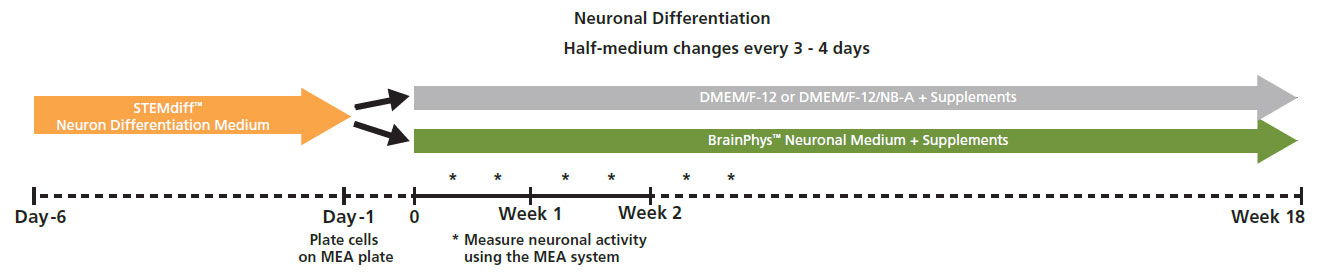
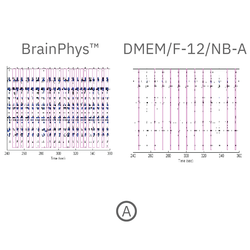
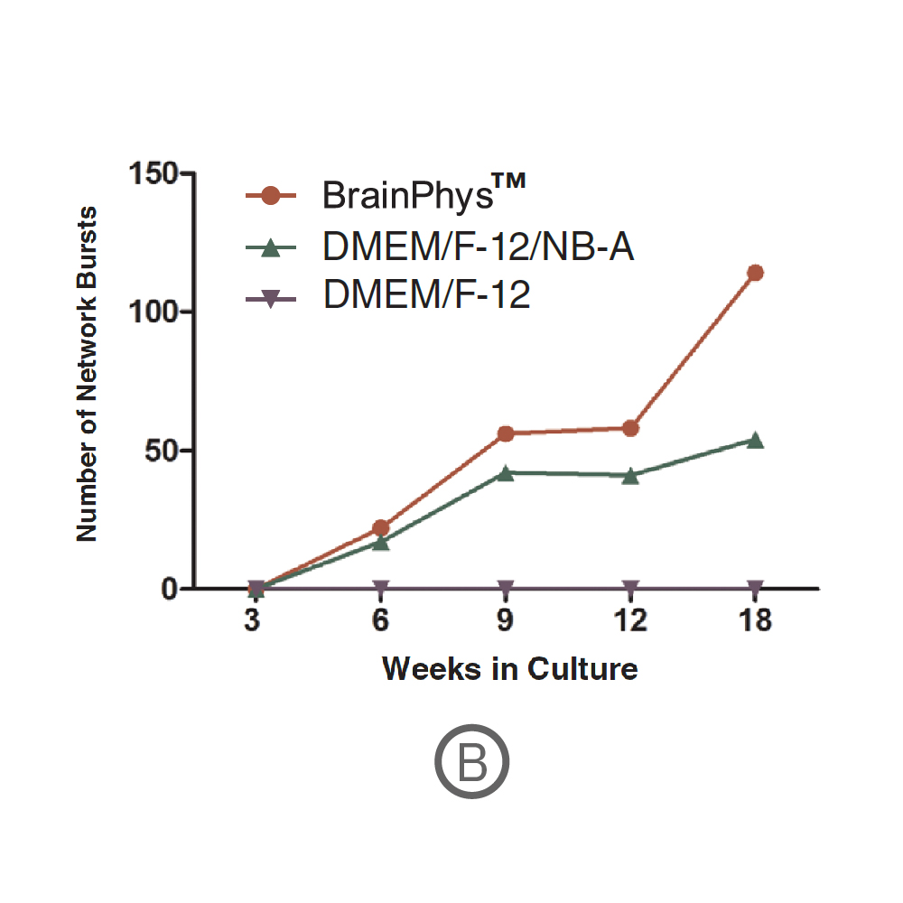
hPSC-derived neurons were monitored longitudinally under different media conditions. Network bursts emerged at week 6 and increased with maturation. At week 18, BrainPhys cultures produced 114 network bursts during a 10-minute recording compared with 54 in DMEM/F-12/NB-A cultures.Data courtesy of STEMCELL Technologies, taken from Mak et al. 2016 presented at SfN2016.
Why functional optimization matters
A culture may look neuronal or express appropriate markers without developing the electrical properties required for the intended experiment.
Functional measurements can help researchers determine:
- when spontaneous activity emerges;
- when networks become synchronized;
- which culture conditions support stronger network activity;
- when the model reaches an appropriate experimental window;
- whether the functional phenotype remains stable over time.
Neural network maturation involves changes at multiple temporal scales.
MEA can record conventional spike activity and local field potentials simultaneously, providing complementary information about individual neuronal firing and coordinated population-level activity.
In the rodent cortical neuron example shown on this page:
- at DIV14, LFP events were small, biphasic, and short;
- by DIV21, oscillatory activity emerged;
- by DIV28, network activity showed stronger initial peaks and delayed rebound events.
These changes reflect increasing complexity as the neuronal network matures.
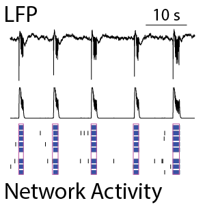
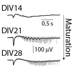
(Left) The raster plot (bottom) identified bursts of spiking detected on individual electrodes (blue) and coordinated bursts of activity across electrodes (pink) for rodent cortical neurons after 21 days in culture. The oscillations in the network activity were detected via in the population activity histogram (middle). The local field potential (LFP) signal (top) was measured simultaneously from each electrode in the well, with one example trace depicted here. The LFP events were coordinated with the network activity in the well. (Right) The detected LFP events (gray) are presented along with the average LFP across events (black) at different stages of neuronal network maturation for rodent cortical neurons. At 14 days in vitro (DIV), the LFP events are small, biphasic, and short in duration. At DIV21, the LFP reveals oscillations in the neuronal network. At 28 days in vitro, the network displayed strong initial peaks with rebound events occurring at variable delays.
Why measure both?
Spike activity shows when neurons are firing.
Network metrics reveal coordination across the culture.
Local field potentials provide additional information about slower population-level oscillations.
Together, these measurements provide a broader functional picture of neural maturation.
Different neural models can develop characteristic patterns of electrical activity based on cell type, composition, and maturation state.
The current page demonstrates this with primary neuronal cultures derived from different regions—including frontal cortex, spinal cord, hippocampus, and midbrain—which displayed distinguishable spontaneous activity patterns after 28 days in vitro.
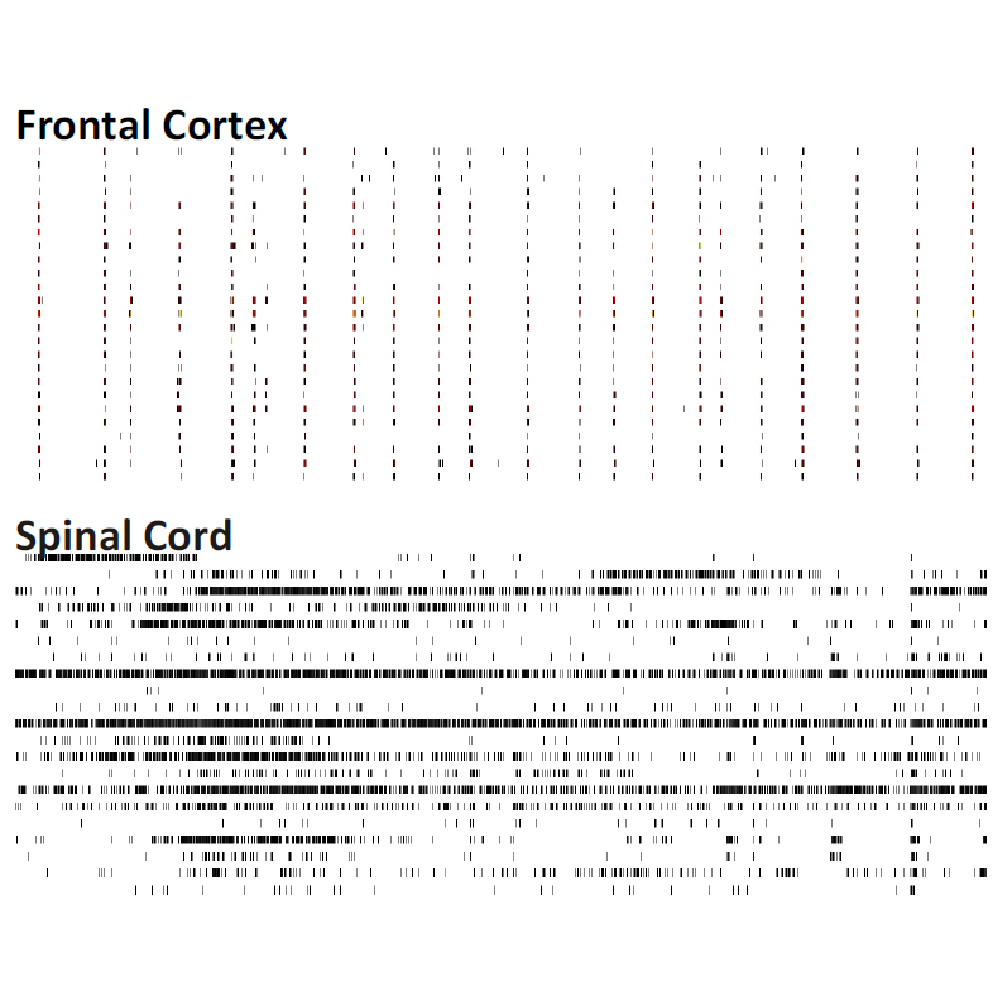
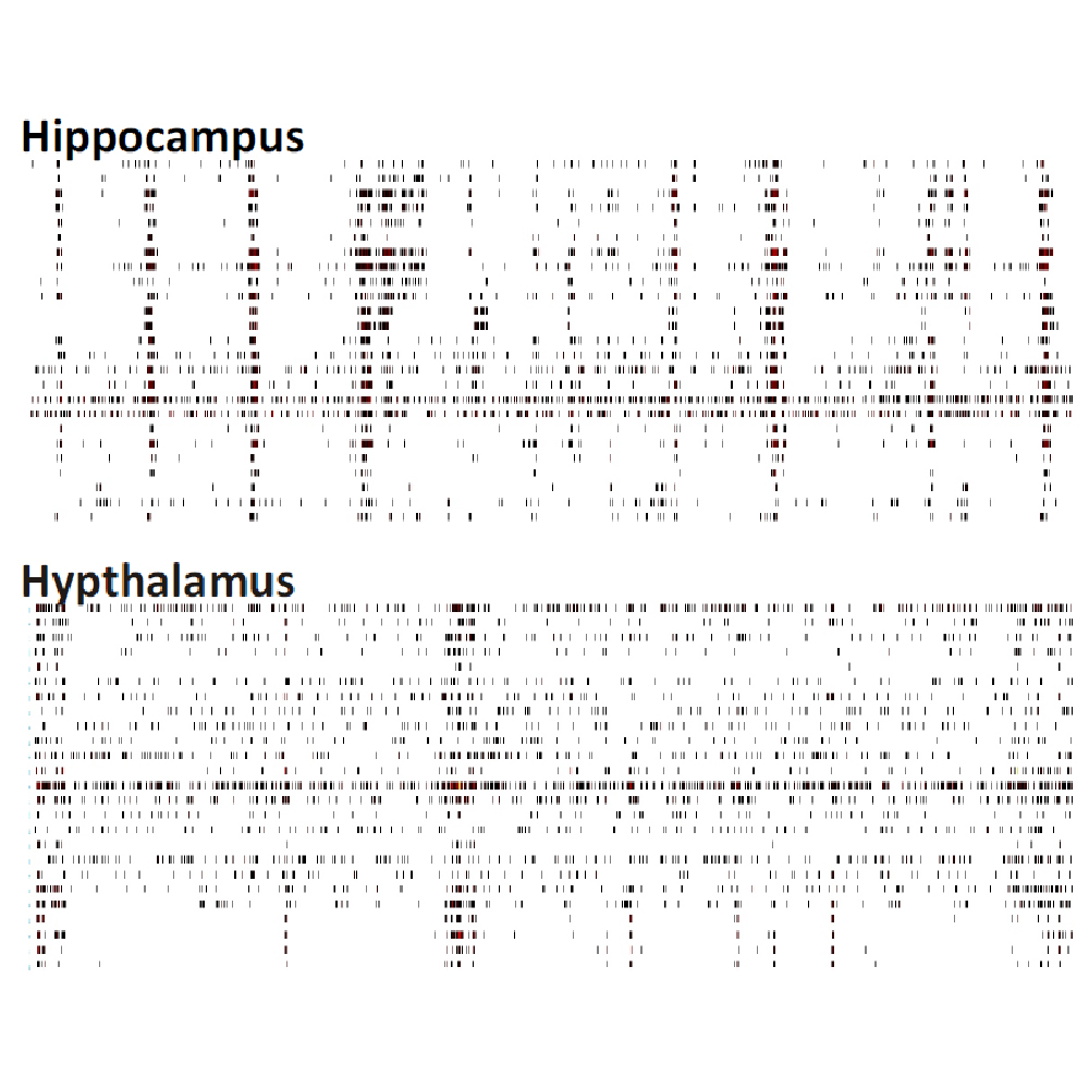
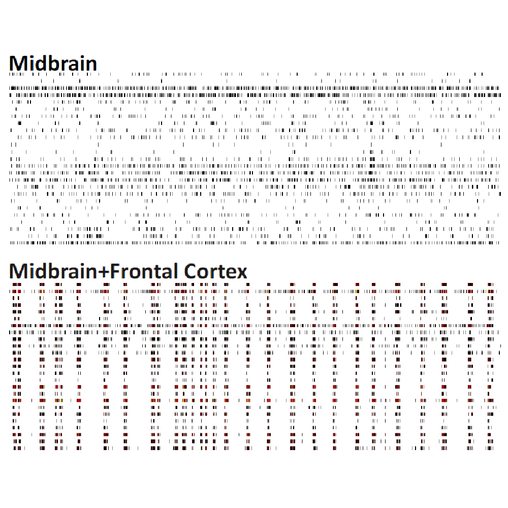
Raster plots of brain region-specific primary cell cultures derived from embryonic murine tissue of the frontal cortex, spinal cord (with dorsal root ganglia), hippocampus, and midbrain (co-cultured with frontal cortex) were compared. Plotted are 60 seconds of 25 neurons of spontaneous network activity at 28 days in vitro. Spontaneous activity plotted for 60 seconds shows distinguishable patterns based on the brain region of origin. Data courtesy of Neuroproof GMBH, taken from Voss et al. 2014 presented at SfN2014.
For stem cell researchers, the same principle can be applied when comparing:
- different neuronal differentiation protocols;
- excitatory and inhibitory neural populations;
- disease and control cell lines;
- different iPSC donors;
- cocultures;
- engineered neural models.
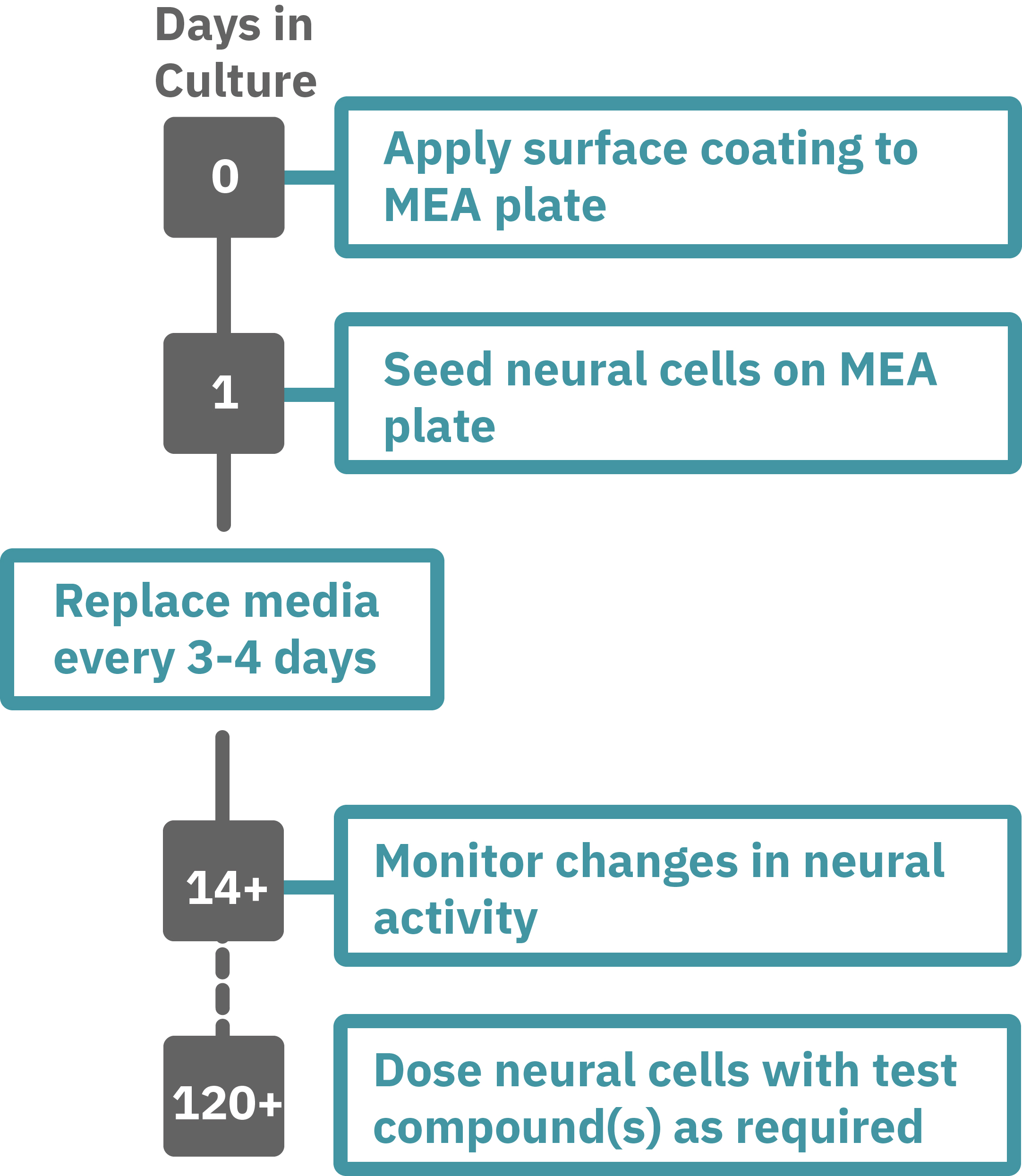
A typical MEA workflow can include:
- Differentiate neural progenitor cells.
- Plate cells on a coated MEA plate.
- Establish spontaneous electrical activity.
- Record repeatedly as the network matures.
- Quantify firing, bursting, synchrony, and other functional metrics.
- Compare developmental trajectories across experimental conditions.
Protocol taken from Mak et al. 2016 presented at SfN2016.
Neural progenitor cells derived from hPSCs (XCL1-NPC) were cultured in STEMdiff™ Neuron Differentiation Medium on poly-L-ornithine (PLO)/laminin-coated 6-well plate for 5 days.
On day 5, neural progenitor cells were dissociated and single cells were re-plated onto a PLO/laminin-coated CytoView MEA plates at 30,000 cells/cm2 in STEMdiff™ Neuron Differentiation Medium.
After one day, half of the medium was replaced with differentiation media (BrainPhys™ Neuronal Medium + supplements: 1% N2 Supplement-A, 2% NeuroCult™ SM1 Neuronal Supplement, 20 ng/mL GDNF, 20 ng/mL BDNF, 1 mM db-cAMP and 200 nM Ascorbic Acid).
Half-medium changes were performed every 3 - 4 days throughout the culture period.
Spontaneous neuronal activity was acquired at 37°C under a 5% CO₂ atmosphere using the Maestro MEA system. A 15-minute recording was taken twice a week and analyzed with AxIS Navigator Neural Module software.
Research questions MEA can help answer
Has my neural model developed a functional phenotype?
Determine whether stem cell-derived neurons exhibit sufficient firing, bursting, and network organization for the intended experiment.
When does the network become functionally mature?
Use repeated measurements to identify when synchronized activity and more complex network behavior emerge.
Which differentiation or culture conditions perform best?
Compare media, plating density, maturation time, supplements, or other variables using quantitative functional endpoints.
Is the phenotype reproducible?
Track consistency across wells, differentiation batches, and experimental runs.
Featured resources for stem cell-derived neural characterization
Best Practices for In Vitro Neural Assays on the Maestro MEA System
Application Note
Review how cell density, culture duration, media, recording parameters, and other factors influence neural assay performance.
Characterization of Functional Neuronal Activity In Vitro with Local Field Potential Signals
Application Note
Explore how LFP power and burst-associated oscillatory activity change during neuronal maturation and following pharmacological manipulation.
Characterization of in vitro cortical networks and their responses to neuroactive compounds
Application Note
Learn how MEA measures the electrical activity of neural cultures in a high-throughput manner and provides quantifiable endpoints as descriptors of the neural network.
Neural Metric Definitions
Technical Resource
Review definitions of firing, bursting, network, synchrony, and related MEA metrics used to characterize neural function.
Looking for additional methods or applications?
Frequently asked questions
Yes. Human pluripotent stem cell-derived neurons can be cultured on MEA plates and recorded repeatedly as functional networks develop. The current page includes an 18-week hPSC-derived neuronal maturation study.
Common endpoints include firing rate, bursting, network bursting, synchrony, oscillatory activity, active electrodes, and local field potentials. [link to neural metrics document]
Imaging and molecular assays provide information about cell identity and structure. MEA provides a direct measurement of electrical function and network behavior.
Yes. Longitudinal MEA measurements can reveal how culture media, differentiation conditions, plating parameters, and maturation time affect neuronal activity and network development.
Because recording is noninvasive, the same cultures can be measured repeatedly over extended periods. The hPSC-derived neuron study on this page followed network activity for 18 weeks.
In the field, Axion MEA users have reported routinely keeping cells in culture for weeks-to-months and some have kept cultures maintained for over a year.
A local field potential is a low-frequency extracellular signal that reflects coordinated population-level electrical activity. LFPs can be recorded simultaneously with neural spike activity and provide complementary information about network maturation.
For Research Use Only. Not for use in diagnostic procedures.