Norrie JL, Nityanandam A, Lai K, Chen X, Wilson M, Stewart E, Griffiths L, Jin H, Wu G, Orr B, Tran Q, Allen S, Reilly C, Zhou X, Zhang J, Newman K, Johnson D, Brennan R, Dyer MA.
Nature Communications, 2021
A retinal organoid model of human retinoblastoma uncovers new information about cellular identity and may enable preclinical testing of therapeutics
Retinoblastoma is a rare childhood cancer of the developing retina that typically arises due to a germline mutation in the RB1 tumor suppressor gene. Scientists using genetically engineered murine models to study human retinoblastoma have been limited by differences across species, but research published in Nature Communications demonstrates that retinal organoids made from patient-derived induced pluripotent stem cells (iPSCs) have molecular, cellular, and genomic features indistinguishable from human retinoblastoma—findings that provide a model of human cancer which contributes to the understanding of the cellular origins of retinoblastoma and may enable preclinical testing of therapeutic combinations for individual patients.
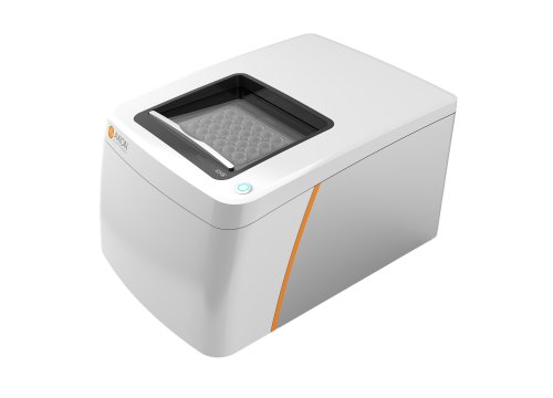
Researchers created the human retinoblastoma model from 15 participants with germline RB1 mutations or deletion and characterized the resulting iPSCs for embryoid body formation, neural rosette formation, and retinal differentiation. The study also investigated RB1 wild-type stem cell-derived retinal organoids as a negative control and induced RB1 mutations with CRISPR-Cas9 in all 15 participant-derived iPSC lines and H9 cells as a positive control. To assess evoked response to optical stimulation in the retinal organoids, the authors used Axion’s Maestro Edge platform with the Lumos multiwell optical stimulation system. Taken together with other results, the findings reveal new information about the cellular identity within retinoblastoma showing a bias toward retinal progenitor cells and rods and demonstrate an optimized 3D retinal organoid culture system for producing human retinoblastomas in vitro.


