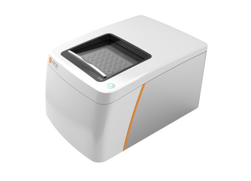
>> Begin with network and spike analysis
Get an in-depth look at your 3D models and discover new insights into brain development, disease modeling, and therapeutic development.
Functionally characterize network and spike activity with an easy, noninvasive assay on the Maestro MEA platform.
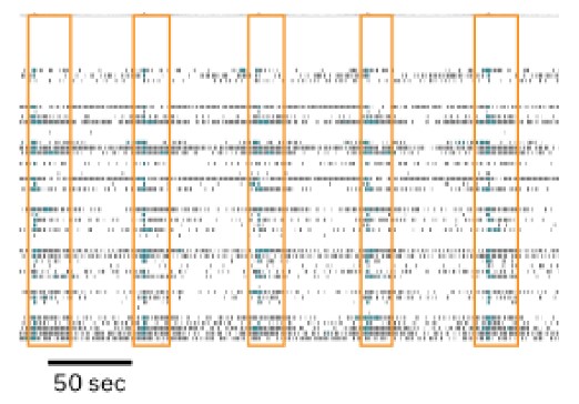
Raster plot of organoid spike activity identifies network (orange) and electrode (teal) bursting.
>> Dive deeper with LFP
Neural organoids can develop complex low-frequency oscillations reminiscent of in vivo brain rhythms. Enhance your characterization and probe your models deeper with local field potentials and power spectral density analysis using Maestro MEA.
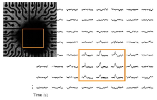
Local field potential (LFP) signals represent the collective activity of the organoid and can be measured simultaneously to the spike activity.
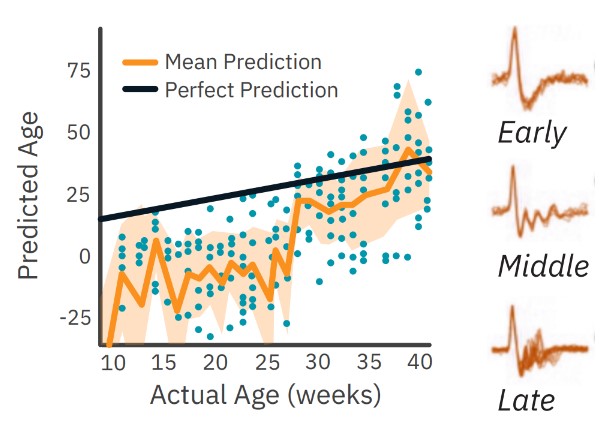
Data from: Muotri, Alysson. (2019, Sep. 17). Measuring oscillatory waves in cerebral organoids [Webinar]. Axion BioSystems.
Track organoid development and maturation
The development of neural networks in organoids resemble those found during early human brain formation. Local field potential (LFP) signals become increasingly complex as the cerebral organoids mature. Researchers in the Muotri lab (UC San Diego) demonstrated that mature organoid activity correlated to in vivo EEG recordings.
>> How it works

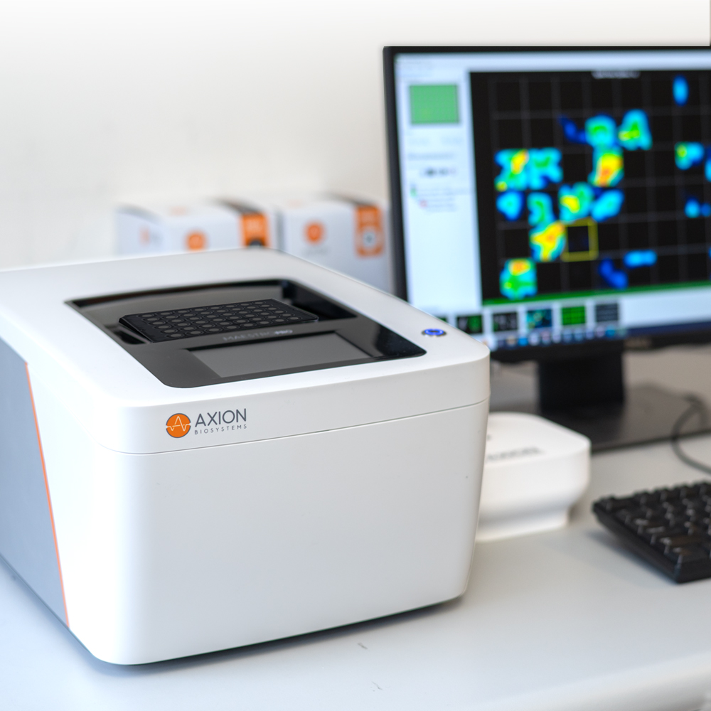
Plate the organoids over the electrodes, confirm attachment with the MEA Viability Module, then record signals on the Maestro MEA. Monitor your organoids as often as you want, for as long as you want. It’s the easy way to get detailed data.
>> Know your organoid is where you want it
We developed the SpheroGuide MEA Plate to make positioning organoids over the electrodes even easier. An integrated plating funnel ensures organoids land where you want them. While the transparent bottom makes it easy to see your organoid, Maestro MEA can also quantify how well it is attached and where to expect signal.
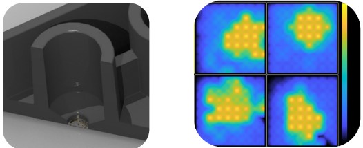
a) Crossection of the SpheroGuide MEA placement funnel. b) Organoid attachment measured by the MEA Viability Module
Frequently Asked Questions
>> How have researchers used Maestro MEA for neural organoids?
There are over 50 peer-reviewed publications using the Maestro MEA for neural organoids. Visit: axionbiosystems.com/neural-organoid-insights for publications, webinars, and more.
>> How do I place an organoid on the electrodes?
Organoids can be placed manually with a pipette or, for higher throughputs, we created the SpheroGuide MEA plate with an integrated placement funnel for fast, easy placement.
>> How do I attach an organoid to the electrodes?
Surface coatings such as PEI or fibronectin can be used to attach the organoid to the bottom of the well. Detailed plating protocols can be found at axionbiosystems.com/resources.
>> Are 3D electrodes required to measure organoid activity?
No, surface cells have activity as well. Nanopillar style electrodes measure just a few cell layers inside the organoid where cells typically have similar activity to surface cells.

