Epilepsy is a devastating neurological disease caused by an imbalance in the electrical signals between the cells of the brain. It affects 1% of newborns and very young children. In children, 1/3 of all cases fail to respond to currently available medication. The primary challenge in the fight against epilepsy is the lack of model systems that effectively mirror what goes wrong in human patients. In this webinar, Dr. Evangelos Kiskinis (Northwestern University), demonstrates how patient-specific neurons in vitro recapitulate the neural firing patterns observed in patient EEGs in vivo. Using this platform Dr. Kiskinis hopes to be able to predict what drug would work best for each patient, removing the trial-and-error approach to epilepsy therapy.
Transcript of webinar on Modeling Pediatric Epilepsy with iPSC-based Technologies
Thank you for joining for today's Coffee Break Webinar Today's topic is: Modeling Pediatric Epilepsy with iPSC-based technologies
Epilepsy is a devastating neurological disease caused by an imbalance in the electrical signals between the cells of the brain. It affects 1% of newborns and very young children. In children, 1/3 of all cases fail to respond to currently available medication. The seizures can result in long-lasting delays in learning, thinking, and emotional development, and even early death. Doctors often have to resort to a trial-and-error approach because it is hard to predict how a patient will respond to a particular therapy. This can have devastating implications for young children and their families.
The primary challenge in the fight against epilepsy is the lack of model systems that effectively mirror what goes wrong in human patients. Scientists have relied on using non-human cells or animal models of the disease, but fundamental differences exist between humans and animals in the biology of the molecules that are implicated in epilepsy.
To solve these problems, Dr. Kiskinis’ lab generates patient-specific stem cells and differentiates these into patient-specific brain cells. By studying their electrical patterns, and how they respond to drugs, Dr. Kiskinis’ hopes to be able to predict what drug would work best for each patient. Removing the trial-and-error approach to epilepsy therapies would have a major impact on how we diagnose and treat epileptic patients.
Dr. Kiskinis is an Assistant Professor of Neurology at Northwestern University Feinberg School of Medicine. He also serves as the Scientific Director of the Stem Cell Core Facility and is the Co-Director of the Stem Cell and Regenerative Biology Initiative. His lab aims to identify points of targeted and effective therapies for epilepsy and ALS by using human pluripotent stem cells.
Thank you for the introduction, Melissa.
Each year about 1 in 100 children are diagnosed with a form of pediatric epilepsy In the most severe cases, babies start seizing within hours, or days after they are born. The seizures, which result from aberrant electrical activity in the brain, are multiform, relentless and exceptionally hard to treat. As a result these kids suffer serious comorbidities including cognitive, behavioral and neurological deficits.
Clinicians have no effective way of selecting the right anti-epileptic drug (AED) for each individual and often resort to a trial and error approach, which can have detrimental effects for babies that are in developmentally very sensitive stages. Pediatric epilepsy, which is what we are interested in, has extensive genetic diversity with 50% caused by de novo mutations in genes that encode ion channel proteins, which control neuronal excitability. These include sodium channels and potassium channels, and in my lab we have been developing programs looking at a number of these variants using iPSC-based approaches.
I believe that iPSC-based technologies are very well suited for the study of pediatric epilepsy for a number of reasons. It is really hard to create animal models that accurately recapitulate the human condition because a) several ion channels exhibit human-specific expression patterns b) clinical severity for these syndromes depends on the interaction of each genetic variant with the patient-specific genetic background.
Now, iPSC models retain the genetic background of patients and allow us to examine distinct neuronal subtypes, breaking down a complicated system like the brain in smaller parts. iPSC differentiated neurons resemble neurons of late embryonic, early postnatal stages, which is the time at which seizures begin for this syndrome. Most importantly, these models allow us to determine the effects of variants on neurodevelopment and excitability and evaluate the ability of small molecules to reverse or slow down these defects.
Now, our goals are to develop biologically relevant and screen-able models of epilepsy, and use these to stratify patients for optimal drug selection. I will use our work on KCNQ2-associated epilepsy as proof of principle to illustrate the range of scalable assays we developed in order to improve drug selection for these kids. The KCNQ2 gene encodes a 6-trans-membrane domain protein that is responsible for neuronal M-current.
There are currently more than 300 distinct variants associated with KCNQ2-epilepsy and clinically they all are treated in the same way. The 1st step in our analysis platform is a high-throughput functional assay that combines heterologous expression of either the wild type, or mutant protein, with automated whole-cell patch instrumentation. This is an example of the primary data, with the activity of the WT or a mutant protein.
This system allows us to collect measurements from hundreds of cells and screen through a high number of variants in a relatively short amount of time. We use these functional data to classify variants based on their pathogenic effect on the channel: some are benign, some cause severe loss-of-function, some mild and some cause increased activity. We then select representative variants from each one of these groups and analyze their effects in patient neurons using iPSC-based technologies to gain further mechanistic insight on how these variants impact neural development and function.
This is the 2nd step in our analysis platform. Here is an example of 2 patient mutations classified with severe loss of function. We generated patient specific induced pluripotent stem cells and then used these to differentiate cortical excitatory glutamaturgic neurons.
One of the assays that we developed allows to measure functional M-current i.e. the function of this protein (something never done before in human neurons). We do this by a whole cell voltage clamp in combination with pharmacological inhibition. We find that while the density of current is consistent amongst neurons derived from 3 unrelated healthy control individuals it exhibits a robust reduction amounting to 55% in neurons derived from patient 01. Importantly correcting the mutation using CRISPR, restores the protein function to a level that is equal to control neurons, while the 2nd patient mutation showed a similar reduction.
These findings are really exciting for us as they provide us with a human neuronal platform to screen for effective channel agonists, which have been proposed as therapeutics for these patients.
We also have been developing assays to measure the functional outcome of these mutations on neuronal firing. Children with KCNQ2-associated epilepsy exhibit a characteristic pattern in their brain EEG known as a burst-suppression activity pattern. They specifically show highly synchronized firing, referred to as bursting, followed by intermittent periods of very little activity.
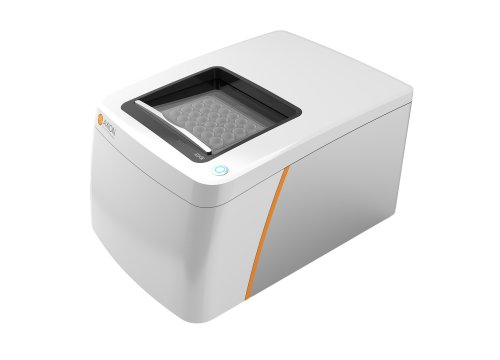
To assess the firing patterns of iPSC-derived neuronal populations we use multielectrode arrays, culturing neurons on fitted electrodes and recording spontaneous firing over several weeks in culture. We find that patient neurons exhibit a significant and time-dependent increase in their firing rates, with a higher number and percentage of synchronized bursts.
In fact representation of the iPSC-derived neuron data in a raster plot, highlights a bust-suppression firing pattern that is highly reminiscent to the one seen in the brain EEG of the same patient, Effectively recapitulating “epilepsy-in-a-dish” and providing us with another phenotypic platform that can be used to assess potential therapeutic options.
We have also established an optical physiology assay that enables high-throughput phenotyping of thousands of patient neurons This is referred to as the Optopatch. It relies on the co-expression of 2 proteins: a channel rhodopsin that triggers neural activity in response to blue light, and an arch rhodopsin that emits precise amounts of red light in response to neural activity. This movie shows neurons expressing theses constructs firing spontaneously and in response to stimulation. By collecting the red fluorescence signal and applying image analytics tools, we record various excitability features with single cell resolution. On the right is the recording from approximately 3 thousand patient and isogenic control neurons under a blue light step protocol, where we gradually increased the stimulation intensity. At the bottom is the aggregate data which shows that the patient neurons exhibit increased firing frequency under all conditions tested. With this technology we capture a number of excitability features represented in this radar plot such as frequency and spike width. The red line represents the average properties of patient neurons and the blue line the properties of isogenic control neurons.
We can also measure the ability of small molecules to restore these defects. As an example when we apply a KCNQ2 channel agonist, we can rescue of number of defective neuronal features such as frequency and adaptation, while others that represent dyshomeostatic secondary effects of channel dysfunction are unaltered.
In summary I have illustrated how we have been developing tools and assays with the goal of impacting pediatric epilepsy research and treatment, a disease that has been largely overlooked and is particularly amenable to modeling with iPSC-based technologies. I believe that this will be invaluable to the community as the entirety of what we know about these syndromes is based on limited non-human based model systems
Here is where I see this work in 5 years: Firstly we aim to analyze as many as 1000 epilepsy-associated genetic variants and classify them based on their effects on function . Secondly we will provide mechanistic insight into how at least 50 representative variants impact human neuronal subtypes using a range of approaches. Thirdly we will determine whether iPSC models can be used to select patient-specific drugs, by performing drug treatment experiments on patient derived neurons.
In collaboration with neuro-geneticists and computational biologists we will integrate our datasets to stratify pediatric epilepsy cases. Our goal is to enable clinicians to rationally and effectively treat these kids and push through precision care in epilepsy.
The work that I presented today was done in collaboration between my lab and Al George’s lab In the Department of Pharmacology here at Northwestern University And was spearheaded by Dr. Dina Simkin and Dr. Tim Searl. We also collaborated with Carlos Vanoye and John Millichap who provided all the primary patient samples which all our iPSC cell lines were generated. I also want to mention that all the Optopatch-based work is in collaboration with Luis Williams and Graham Dempsey and Owen McManus at Q-State Biosciences I also want to thank our funding agencies that have supported this work.
And that is the conclusion for today’s Coffee break webinar. If you have any questions you would like to ask regarding the research presented or if you are interested in presenting your own research with microelectrode array technology, please forward them to coffeebreak@axionbio.com. For questions submitted for Dr. Kiskinis, he will be in touch with you shortly.
Thank you for joining in on today’s coffee break webinar and we look forward to seeing you again.


