Autism, also known as autism spectrum disorder, is a range of conditions classified as neurodevelopmental disorders. Individuals diagnosed with autism show challenges with social skills, repetitive behaviors, speech and nonverbal communication. Autism is estimated to affect about 1% of people or 62.2 million globally. The genetics of autism are complex meaning better methods are required to help understand the genetic risk factors that underlie autism. In this webinar, Dr. Michael Nestor (The Hussman Institute for Autism) discusses how studying the spontaneous firing activity of patient-derived iPSC neurons in an MEA assay is helping to build a model of autism.
Audio Transcript:
Thank you for joining us on today's coffee break webinar, today's topic is 'the spectrum in a dish' using neurophysiology to build a human IPSC model of autism. Autism, also known as Autism Spectrum Disorder, is a range of conditions classified as neurodevelopmental disorders. Individuals diagnosed with autism show challenges with social skills, repetitive behaviors, speech, and nonverbal communication.
Autism is a spectrum condition - as it affects individuals differently and to varying degrees.
For someone on the high-functioning end, autism may present relatively mild challenges, but for individuals with more severe symptoms repetitive behaviors and lack of spoken language can interfere with everyday life. Symptoms are typically recognized between 1 and 3 years of age though it is still considered a lifelong condition appropriate early intervention can reduce symptoms and increase skills and abilities. Autism is estimated to affect about 1% of people or sixty 2.2 million globally. While specific causes of autism have yet to be found, there is a strong genetic basis to the disorder. The genetics of autism are complex, meaning better methods are required to help understand the genetic risk factors that underlie autism.
Today's presenter Dr. Michael Nester, is an investigator at the Hussman Institute for autism and the co-chair of the Neural Stem Cell Working Group at the University of Maryland School of Medicine. He is a neurophysiologist with 15 years of experience and a focus on electrophysiology, neural stem cell biology, and project management. In particular, his laboratory works on assay development with an emphasis on cell-based preclinical high content drug screens, and phenotyping assays involving both 2D and 3D human iPSC-derived neurons from individuals with autism. He will discuss how studying the spontaneous firing activity of patient-derived iPSC neurons in an MEA assay is helping to build a model of autism.
Hello, my name is Michael Nestor, today I'm going to be talking to you about work in my lab using neurophysiology combined with human iPSC-derived neurons to build a model of autism. The Hussman Institute is a new Institute in Baltimore Maryland, we're focused solely on enhancing the lives of individuals with autism, and to do this we have both clinical and basic research facilities. On the basic research side we focus on core research areas in neuroscience related to things like GABAergic circuitry, excitatory-inhibitory balance, cell adhesion neuroanatomy, cell culture, and induced pluripotent stem cells, amongst others, we recommend that you check out our website for more information.
Autism is a condition that affects both individuals and the families that support them, autism affects 1 to 1.5 million people in the United States. Although autism represents a spectrum of behavioral and clinical phenotypes, there are some convergences, particularly in behavioral phenotypes around disruption and, social communication and social interactions. This can include restricted or repetitive behaviors, restricted or repetitive interests, or restricted or repetitive activities. Although in approximately 30% of cases, there's comorbidity with epilepsy, autism can be clustered with a wide range of comorbid conditions reflecting a high level of underlying genetic heterogeneity. In this condition only about 10% of individuals with autism can be clearly connected with underlying chromosomal and genetic disorders, leaving a large area for polygenic and de novo mutations that can contribute to autism in the data.
Today we'll be talking about individuals with idiopathic polygenic autism, and they represent most of the cases of individuals with autism. We're developing platforms for accurately modeling aspects of the excitatory-inhibitory imbalance hypothesis and autism, first put forth by Dr. John Hussman around 2001, and then elaborated by others after that. We're using our developed platforms to perform high content drug screening at our facility at the Hussman Institute for autism as I mentioned earlier we're also asking whether there are points of convergence in the neurophysiological readouts of individuals who have autism but with different genetic backgrounds and it's these points of convergence that we plan to exploit. For drug discovery, although animal models are important and have gotten us to where we are now, autism is a uniquely human condition, and as such animal models are not really adequate to represent the complex underlying biology of autism.
This effectively leaves biologists using animal models operating under a type of observational bias, called the streetlight effect, and you can see that in the cartoon reflected there. Therefore we use human iPSCs derived from peripheral blood mononuclear cells, these are isolated from the blood of individuals with autism and controls. Although we have a large cohort of these individuals the talk today, in the interest of time, will only focus on a small subset of these people. After isolation, which was done by our colleagues at the Hussman Institute for Human Genomics at the University of Miami School of Medicine. The PBMCs were subjected to whole-exome sequencing using the Agilent Sure select system. After sequencing, autism candidates' genes were identified again by our colleagues at the Hussman Institute for Human Genomics. You can get a sense of the protocols and procedures that we use to identify autism-related candidate genes in these PBMCs, by taking a look at the reference in the box on the right-hand side of the slide.
After PBMCs were sequenced they were turned into iPSCs and then iPSCs were turned into human stem cell-derived cortical neurons, using our previously published protocols, and these cortical neurons were made from individuals with autism and controls, in order to model aspects of this condition. I first validated the multielectrode array system is useful for looking at overall network-level effects in iPSC derived neurons, when I worked at the New York Stem Cell Foundation, as I published the first recordings of human IPSC derived dopaminergic neurons, from twins using the Axion system in 2014. The MEA serves as a way to assess overall activity in neural networks that exhibit a large amount of well-to-well and plate-to-plate heterogeneity.
In our case, this heterogeneity is confounded by the fact in autism we are also looking at the genetic heterogeneity of each individual, therefore, in contrast to the obvious constraints of single-cell electrophysiology, the MEA allows for the recordings of large numbers of neurons, across large numbers of individuals to give a more accurate representation of the biology in a dish and the variability of recordings.
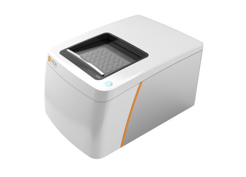
For those who are not familiar with the multielectrode array, multielectrode arrays record local field potentials (LFPs) are the electrical activity of groups of neurons, this is usually induced by the release of neurotransmitters by those neurons. In contrast to single-cell electrophysiology, the LFP and the spikes that are recorded in the multielectrode array are an emergent property of in vitro spontaneously active, functional neuronal networks. They underlie in vivo physiology and they are also tangentially related to the single-cell spike activity of the neurons that surround any given electrode.
Additionally, we found in this cohort of individuals with autism, using both transcriptome analysis and weighted gene co-expression network analysis, that there were significant differences in the pathways related to general neuronal function and synaptic function, and those pathways converged across all the individuals in our cohort. So, this prompted us to perform the MEA recordings in the first place. What you're looking at is spontaneous spiking in a mixed population of iPSC-derived cortical neurons.
In our population, we have about 30 percent inhibitory and 70 percent excitatory neurons. 114 and 574, our controls and 110, 134, 691, 709, 725, and 732 are our individuals with autism, and what you can see if you just look at spontaneous activity, spontaneous MEA recorded spiking activity normalized the controls, that all the individuals with autism had a significant decrease in overall spike activity, except for individual 110 who has no significant difference from controls.
To probe single-cell activity in a high-throughput way, we use somatic calcium transients measured by application of the calcium indicator, Flou-4, as a rough analog for action potential activity that can be measured across the entire dish. We observed a significant decrease in the number of calcium transients in the autism lines, as compared to controls. This reflected the electrophysiological recordings we obtained in the MEA. Interestingly, line 110 which was no different from the controls in the MEA recordings, is different from the controls with respect to single-cell calcium transients. This suggests a divergent molecular pathway in 110 from the other autism lines, at least with respect to calcium signaling.
We are now designing more specific tests to understand what the difference is between the electrophysiological recordings obtained in the MEA for 110 and the single-cell calcium transients recorded using Fluo-4, in this line. I mentioned earlier that our culture system consists of about 30 percent inhibitory cortical neurons and about 70 percent excitatory cortical neurons, therefore we use pharmacology to dissect the network, by applying Picrotoxin to block GABAergic signaling from interneurons and isolate excitatory neurons. We combine the Picrotoxin with either APV or MPQX to block NMDA receptors and AMPA receptors respectively. You can see the effect of that drug cocktail on the overall spike activity in the activity map on the left of the slide, to the right of the slide you can see that only APV had an effect on spike activity, by looking at the local field potentials with PTX and APV application, and the raster plots below the local field potentials. This APV effect on spike activity suggests to us that at least some of the effects we observed in the MEA and the Fluo-4 recordings may be due to the function of NMDA receptor activity in these networks.
Morphologically, we also looked at the number of dendrites from iPSC derived neurons that were able to reinvade a scratch as part of a scratch assay, you can see an example of the scratch on the left-hand side of the screen. We found that after about one week the individuals with autism displayed a significant decrease in the number of processes that grew back into the scratch, you can get a sense of the variability between cases and controls, by looking at the graph in the lower right-hand side of the screen. Now that we've established preliminary phenotypes in 2D iPSC-derived cortical neurons, we are now turning to 3D organoid-like cortical structures called serum-free embryoid bodies, or SFEBs. We have published protocols on how to record SFEBs on multielectrode arrays in order to understand the dynamics of networks, in a more complex system of autism. You can see here that we're able to record spikes from SFEBs on MEAs at dates 30 and 90 and by giving drugs that either disrupt excitation or inhibition, we're able to disrupt the number of continuous spikes recorded from the SFEBs at day 30 or day 90.
Today we've shown you a little bit of data on how we've developed the 3D system for deriving cortical cell types that can be used somewhat interchangeably with traditional 2D cultures, for uncovering phenotypes in autism, we were able to reveal physiological phenotypes in the system using the multielectrode array in the 2D culture system, we have developed physiological assays that are scalable to high-throughput and have already begun to yield some preliminary clues as to a cellular phenotype in the cohort that we've shown you today.
This work is only possible through the contributions of a number of very talented people, I would like to particularly thank our colleagues at the Hussman Institute for Human Genomics for their bioinformatics work and for providing us the iPSCs that we derived into neurons, to develop the assays that we've shown you today. I would also like to give a special thanks to John Hussman, the founder of the Hussman Institute for Autism, and The Hussman Institute for Human Genomics for supporting this work and allowing it to continue, and Gene Blatt the director of Neuroscience at The Hussman Institute for Autism for his support of this work as well, and last but not least to all the families who make this work possible.
Thank you and that is the conclusion for today's coffee break webinar, if you have any questions you would like to ask regarding the research presented or if you are interested in presenting your own research with microelectrode array technology please forward them to coffeebreak@axionbio.com. For questions submitted for Dr. Nester, he will be in touch with you shortly.
Thank you for joining in on today's coffee break webinar and we look forward to seeing you again!


