Authors: Cyrielle Jajkiewicz, PhD, Valérie Pouliot, MSc, and Mohamed Chahine, PhD
HeartRhythm, 18 February 2026
Researchers use the Maestro platform to track progressive electrophysiological dysfunction in patient-derived cardiac organoids modeling myotonic dystrophy type 1.
Myotonic dystrophy type 1 (DM1) is a multisystem disorder that can cause serious cardiac complications, but many in vitro models fail to capture how dysfunction evolves over time. To address this, researchers used developmentally mature, patient-specific 3D cardiac organoids to investigate the temporal progression of DM1-associated cardiac abnormalities, with the goal of modeling disease features that may not emerge in traditional 2D cultures.
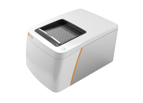
Using Axion BioSystems’ noninvasive Maestro MEA platform, the team recorded electrophysiological activity from both ventricular and atrial cardiac organoids over time. In left ventricular organoids, abnormalities were already evident by day 6, including increased corrected field potential duration (FPDc), prolonged excitation-contraction delay, and elevated beat amplitude. By day 30, these ventricular organoids showed further worsening, with additional decreases in spike amplitude, spike slope, and conduction velocity. In atrial organoids, the disease signature was distinct, with increases in FPDc, spike amplitude, spike slope, and conduction velocity, while excitation-contraction delay remained unchanged.
These findings show that cardiac organoids can capture chamber-specific and time-dependent features of DM1, offering a more physiologically relevant model for studying late-stage disease mechanisms. The work also highlights the value of Maestro-based functional recordings for resolving how cardiac dysfunction emerges and diverges across organoid subtypes.