Understanding in vitro cardiomyocyte characterization is vital for accelerating cardiovascular research, improving drug discovery, and fostering the development of new therapies for heart-related conditions. Characterizing cardiomyocyte function in vitro is critical for studying heart cells, from the initiation of action potentials, propagation across the syncytium, to contraction.
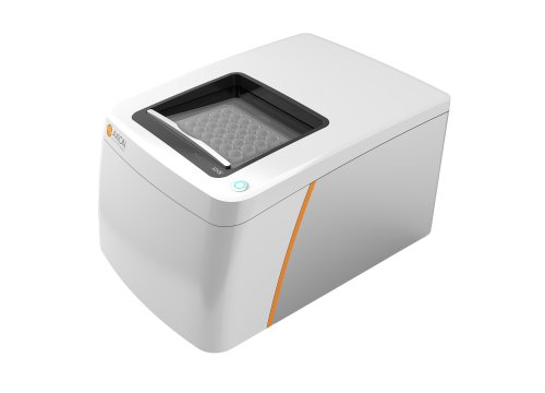
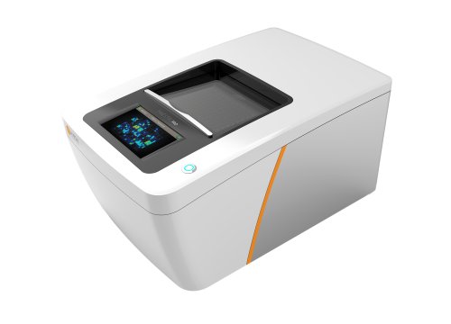
The Maestro MEA platform measures all of these for a deeper understanding of cardiac biology – ideal for both disease research and drug discovery and safety.
Comprehensive in vitro cardiac function assay
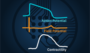
The Maestro MEA platform is more than MEA, measuring both electrical and contractile properties across the culture.
Electrophysiology
- Field & action potential analysis
- Beat & arrhythmia tracking
Contractility
- Contraction amplitude
- EC coupling
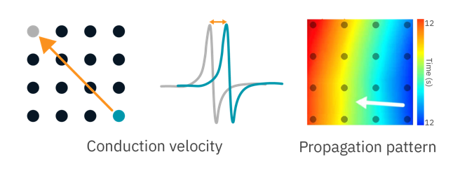
Propagation
- Conduction velocity
- Pattern recognition
Together, you get a more complete functional analysis with one system and one plate.
Reveal hidden mechanisms driving cardiac disease and arrhythmias with the only system offering this comprehensive in vitro cardiac activity assay.

Real-time, label-free in vitro cardiac analysis
-
Measure real-time, label-free in vitro cardiac activity>
-
Detect subtle changes in action potential morphology>
-
Evaluate inotropic effects with contractility>
-
Track propagation across the cardiac syncytium>
-
Highly reproducible and predictive in vitro cardiac activity assay>
-
Establishing a functional standard for stem-derived cardiomyocytes>
-
Elicit mature force-frequency responses with chronic pacing>
-
Cardiac organoids to model ischemia-reperfusion injury>
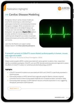
-
Publication Highlights: Cardiac Disease Modeling>
Measure key metrics of cardiac depolarization, repolarization, beating and arrhythmia. Cardiac field potentials can be measured label-free in real time using Maestro MEA. The method is noninvasive and requires only basic cell culturing technique allowing for long-term monitoring of cultures, or acute drug responses.
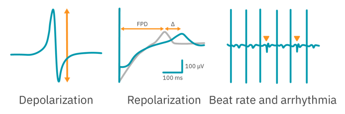
To demonstrate MEA’s ability to characterize cardiac activity, human iPSC-derived cardiomyocytes were dosed with cardioactive compounds.
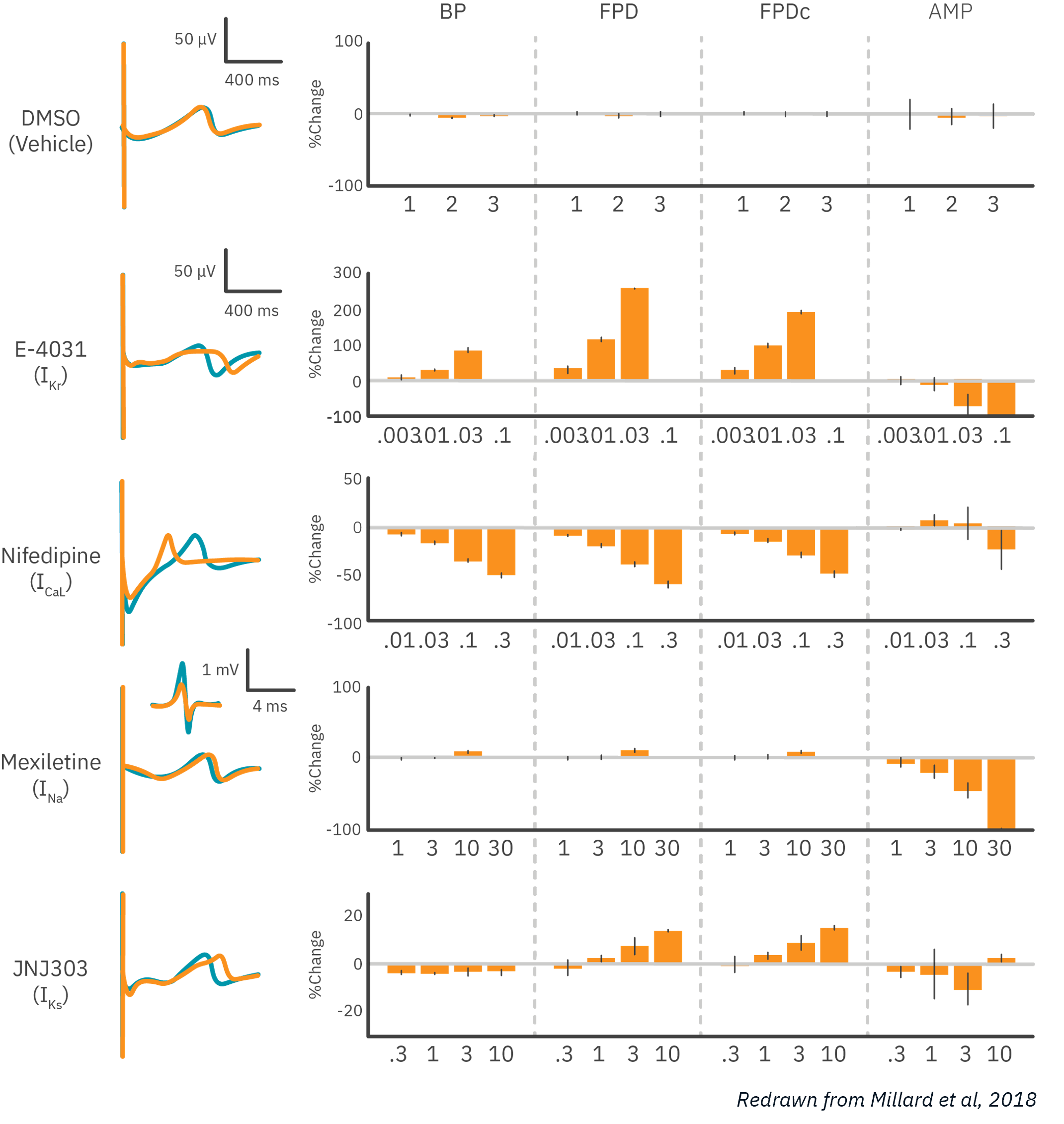
Results: MEA was able to detect dose-dependent effects of compounds targeting major cardiac ion channels. As expected, potassium channel block prolonged field potential duration (FPD), while calcium channel block shortened FPD, and sodium channel block reduced the amplitude of the depolarization. Data courtesy of Millard et al, 2018 and CiPA.
Cardiac action potentials are the culmination of complex interactions that orchestrate the electrical activity of the heart. LEAP (local extracellular action potential), a unique capability of Maestro MEA, can quantify action potential morphology and detect subtle changes to biology with the best signal:noise ratio. Key metrics, such as rise time, action potential duration (APD), triangulation ratio, and percentage of beats with EADs are all automatically detected.
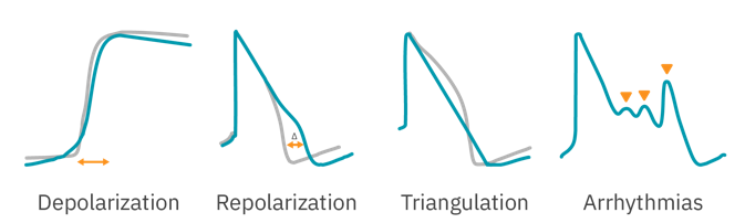
To demonstrate LEAP’s ability to characterize action potential morphology in vitro, human iPSC-derived cardiomyocytes were dosed with cardioactive compounds.
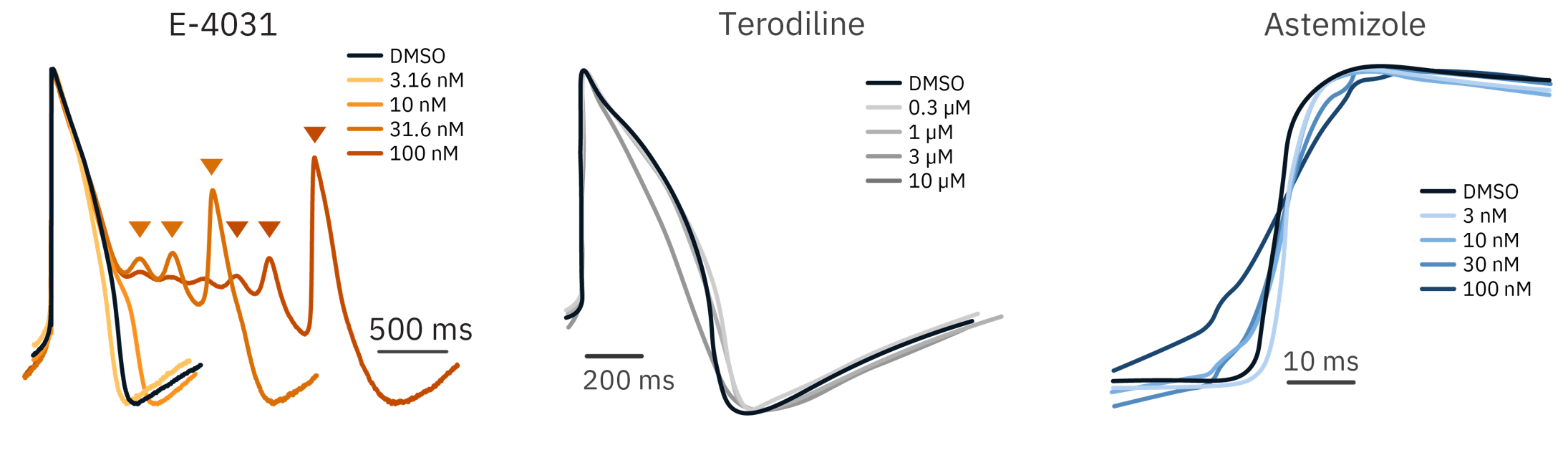
Results: LEAP was able to detect action potential changes indicative of cardiotoxicity: slowing depolarization, waveform triangulation, and early after depolarizations (EADs). Each compound responded in a dose-dependent manner.
Physical contraction is the primary function of the heart. The multimodal Maestro MEA platform can evaluate cardiomyocyte contractility side-by-side its electrophysiology outputs. This enables the quantification of contraction amplitude, beating, and excitation-contraction metrics for the evaluation of inotropic compounds.
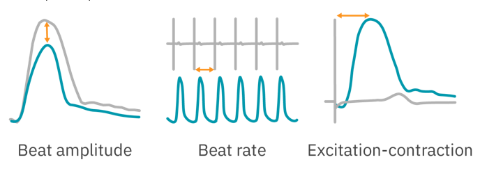
Inotropic drugs can help optimize cardiac output during times of impaired heart function. Here, human iPSC-derived cardiomyocytes were paced for 48 hours before being dosed with cardioactive compounds.
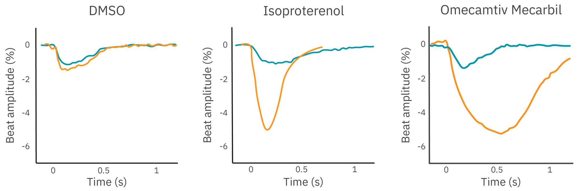
Results: Both beta adrenergic stimulation and myosin activation significantly increased beat amplitude relative to the DMSO control. The Maestro MEA contractility mode is a powerful tool for the investigation of inotropy.
Want to learn more about the inotropy assay?
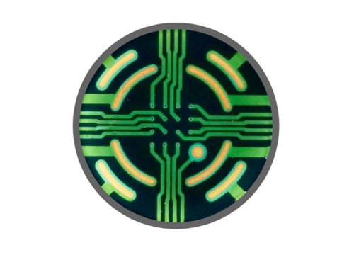
Coordinated propagation is critical to heart function but requires simultaneous measurements from multiple locations on the cardiac syncytium. With an array of sensitive electrodes, Maestro MEA is the ideal tool for assessing conduction velocity and propagation patterns.
To demonstrate the Maestro MEA’s propagation assay, human iPSC-derived cardiomyocytes were dosed with cardioactive compounds.
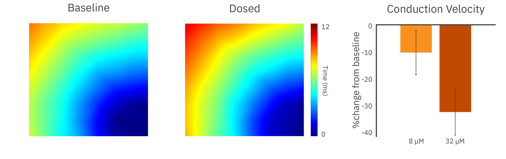
In the presence of a sodium channel blocker, the cardiomyocytes showed a decrease in conduction velocity across the array.
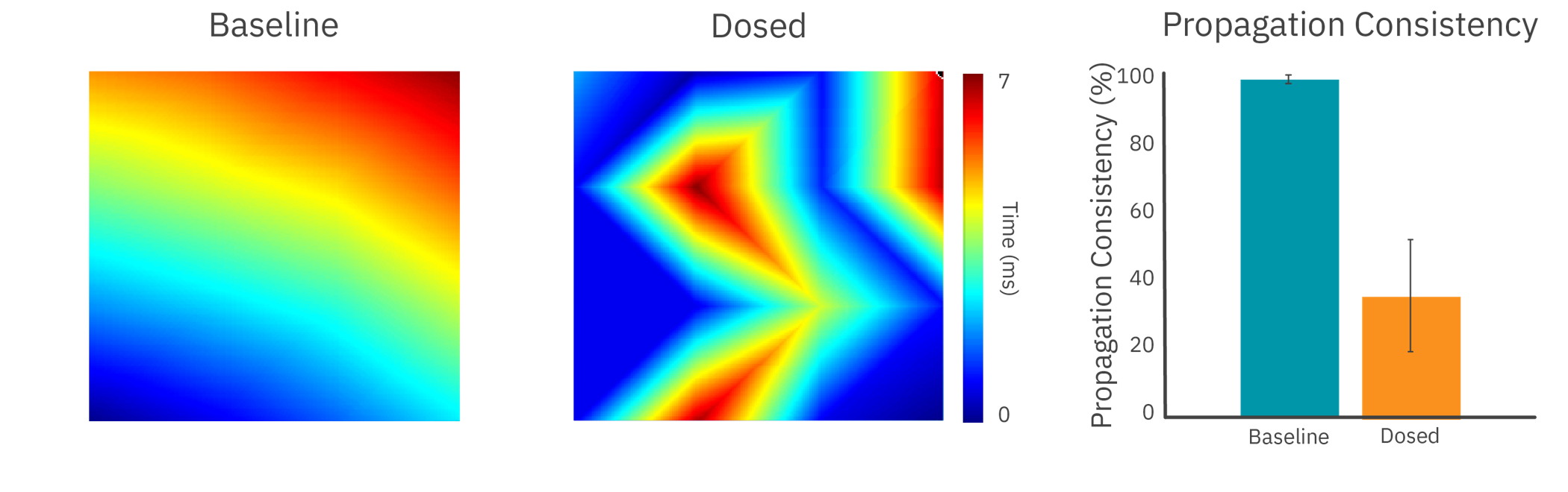
The addition of an anticancer compound disrupted propagation across the syncytium, altering the propagation pattern and reducing propagation consistency.
Results: With high sampling rates across an array of electrodes, Maestro MEA was able to detect slowing and disruption of cardiac propagation across the culture.
The Maestro MEA cardiac activity assay has been proven to be highly accurate with low variability. In a multi-site, blinded study to assess proarrhythmic risk of cardioactive compounds, Maestro MEA outperformed the competition in a head-to-head comparison.
| Maestro MEA | MEA2100 | xCELLigence RTCA CardioECR |
| Most Reliable Sites that passed QC | 85% | 42% | 25% |
| Most Accurate Identified compounds expected to induce a 20% change in FPDc | 93% | 50% | 75% |
| Lowest Variability Well-to-well FPDc variability in baseline | 6.4% | 21.5% | 15.2% |
The study (Millard et al, 2018) investigated 8 compounds in 18 studies across 13 global sites, 3 MEA platforms, and 4 hiPSC cardiomyocyte sources. 85% of Maestro MEA sites passed initial QC, more than double the next MEA system.
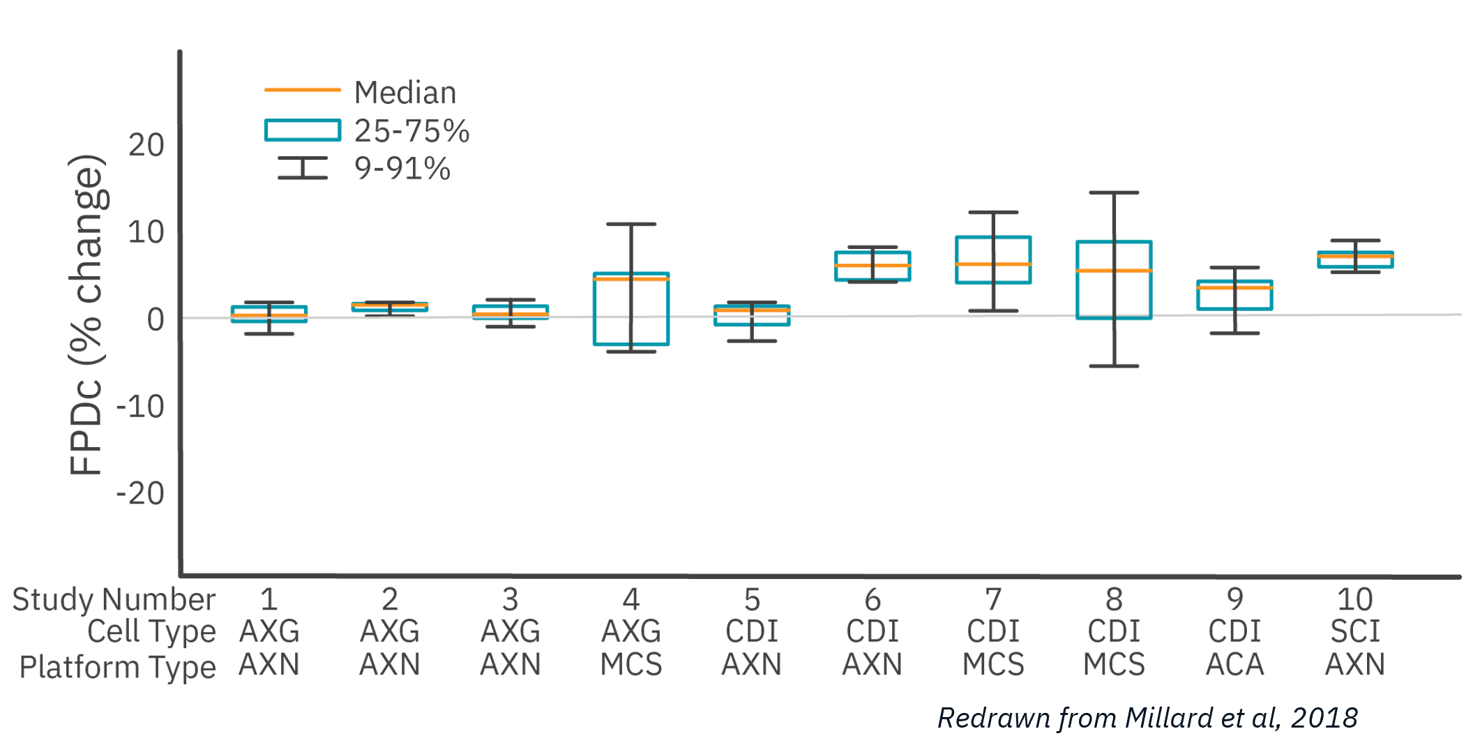
Of the 10 studies that passed QC acceptance criteria, Maestro MEA had the lowest well-to-well variability, 58% less than the next lowest despite being performed across 6X as many sites.
The Maestro MEA assay correctly identified test compounds 93% of the time. A follow-up study (Blinova et al 2018) expanded the scope to 28 blinded compounds of low, intermediate, and high risk. Maestro MEA had a 0.93 correlation coefficient between study sites.
With these studies, together with key opinion leaders in academia and industry, Axion has developed a proposed standard for hiPSC cardiomyocytes.
Together with key opinion leaders in academia and in industry, and combined with published data, we developed a proposed standard for hiPSC cardiomyocyte functionality on MEA. Briefly, this standard focuses on the hiPSC-cardiomyocyte spontaneous beat rate, features of the cardiac waveform (depolarization spike amplitude and field potential duration), and the synchronization of activity in the syncytia.
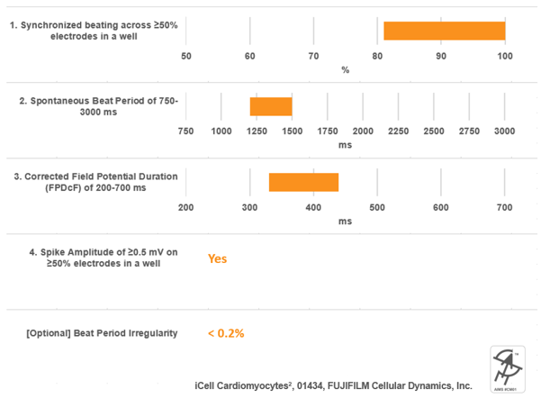
An example of cell performance data from a leading commercial source of hiPSC-cardiomyocytes with respect to this proposed standard.
Want to read the full standard and learn more about Axion iPSC Model Standards (AIMS), including self-certified vendors, recommended protocols, and selected publications?
Purpose: To elicit a mature force-frequency relationship in hiPSC-derived cardiomyocytes. In a mature cardiomyocyte, an increase in frequency is accompanied by an increase in contractile strength. This force-frequency relationship is often lacking in hiPSC-derived cardiomyocytes and is a sign of their immaturity.
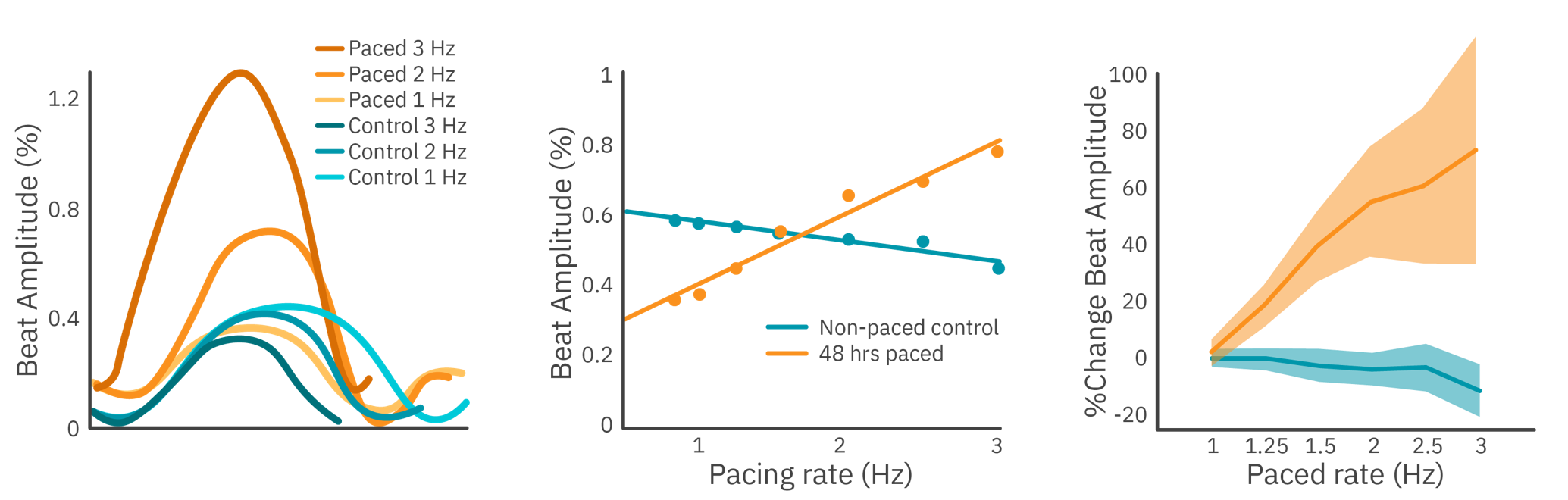
Electrical stimulation was used to measure the force-frequency relationship of hiPSC-derived cardiomyocytes with the Maestro MEA. Prior to testing, the culture was kept on the Maestro MEA for 48 hours, chronically pacing half of the wells.
Results: Cardiomyocytes chronically paced for 2 hours demonstrated a mature force-frequency relationship as measured by beat amplitude using Maestro contractility.
Want to learn how?
Ischemia-reperfusion injury, a primary driver of cardiac disease, is known to affect more than cardiomyocytes alone. Multicellular cardiac organoids provide a more clinically-relevant in vitro model to investigate the complex interactions between the cells of the heart and their implications in disease.
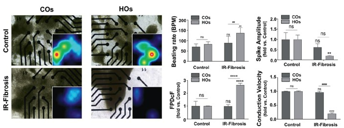
To demonstrate the importance of multicellular interactions in ischemia-reperfusion injury in vitro, and the electrophysiological defects associated with it, cardiac organoids (COs) comprised of cardiomyocytes alone and heart organoids (HOs) comprised of cardiomyocytes, fibroblasts, and endothelial cells were created. Maestro MEA was used to measure activity following ischemia-reperfusion injury.
Results: 3D multicellular heart organoids were an effective model of ischemia-reperfusion injury. Both cardiac organoids and heart organoids led to electrophysiological alterations, but multicellular heart organoids demonstrated a far more robust response. A two-fold increase in field potential duration (Fridericia-corrected) and a significant slowing of conduction velocity was present in the heart organoids, indicative of an elevated risk of arrhythmogenesis. Data from Song et al, 2024, provided by NEXEL.
Publication Highlights: Cardiac Disease Modeling
Review the latest cardiac disease modeling research using Axion’s platforms.

Cardiac Metric Definitions
Discover our Cardiac Metrics Definition guide to discover the full list of metrics available in our cardiac analysis tool.