The brain is a collection of individual, but interconnected functional circuits. Additionally, circuits within the nervous system interact with various other biological systems in the human body. Compartmentalized in vitro models allow two spatially distinct neural circuits or, more generally, two cell populations to develop functional connections and mimic key biological interactions.
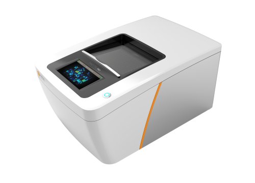
Using Axion BioSystems' Maestro Pro and Edge systems, any scientist can now track functional network formation between neural co-cultures in the same well. Together, neural co-cultures on MEA plates create an ideal model for studying the functional connection between two networks, and the effects of synaptic blockade.
-
Tracking functional network formation between two neural populations>
-
Model neuronal subtype interactions>
-
Isolate and measure neurite activity>
-
Assay Steps>
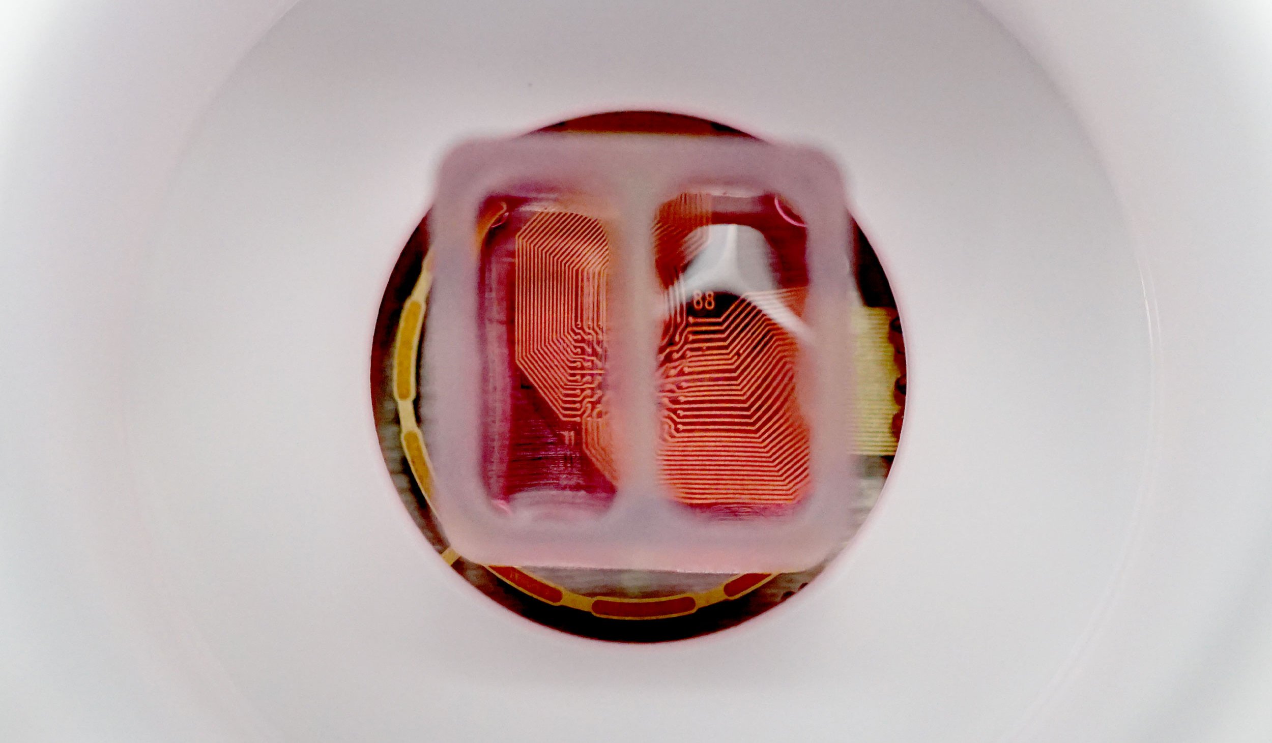
A Culture-Insert 2 Well (ibidi, 80209) composed of silicone was used to culture two cortical neuron populations within the same well of a CytoView MEA 6-well plate (M384-tMEA-6W). At 7 days in vitro, the Culture-Insert 2 Well was removed, such that neurites could cross the cell-free gap and establish functional connections between the two distinct cortical networks. Experimental results from an optical image and activity map confirmed that a consistent, bi-directional functional connection between the two networks was formed within 10 days of Culture-Insert 2 Well removal.
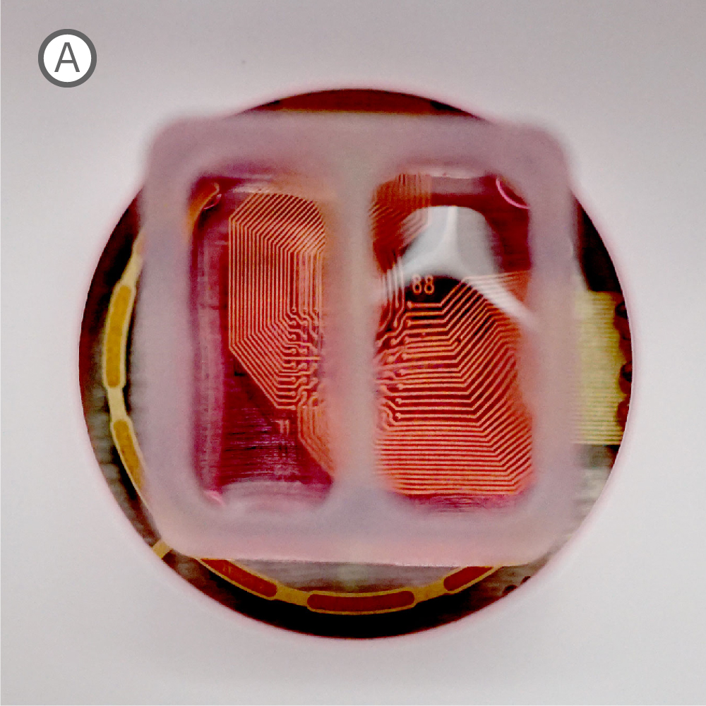
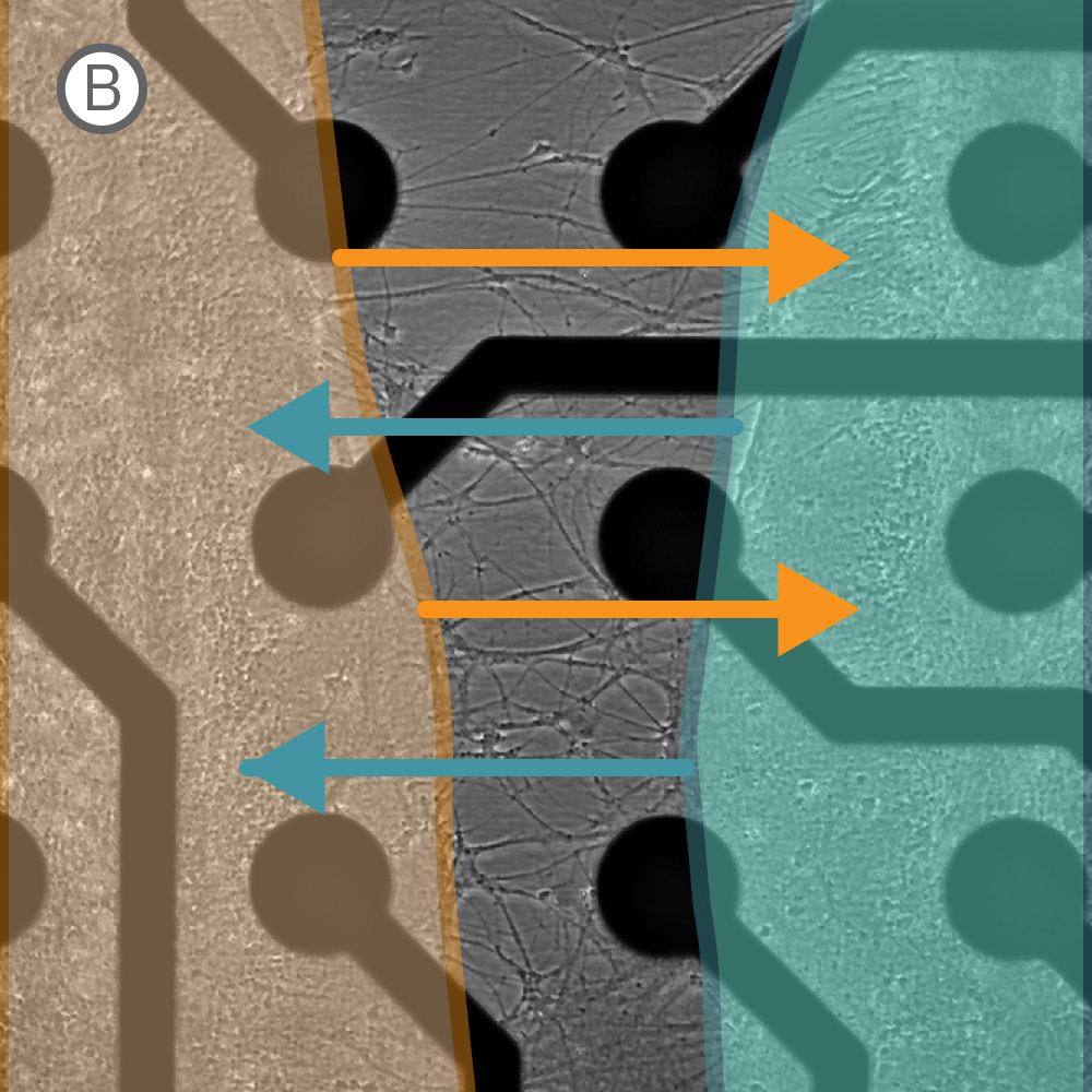
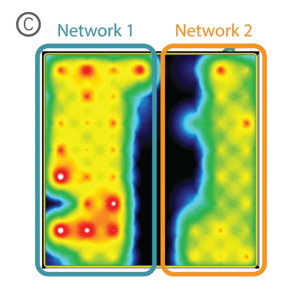
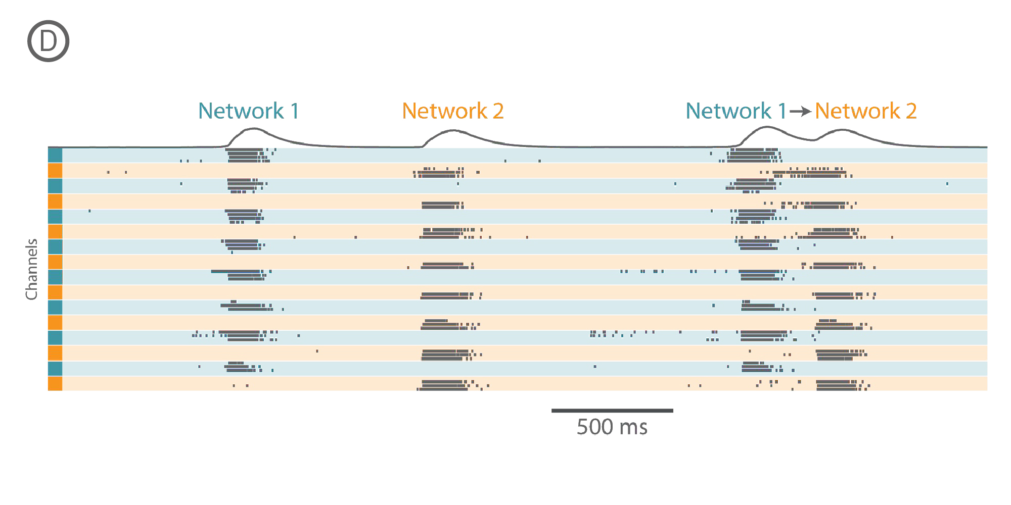
A) Culture-Insert 2 Well (ibidi, 80209). B) Neurites project across the cell-free gap within 10 days of removing the insert. C) Activity map illustrating synchronous activity between the two spatially separated networks. D) Raster plot at 17 days in vitro illustrating independent network activity from Network 1 and Network 2, followed by a whole-well network event with Network 1 driving the activity in Network 2.
Purpose: To demonstrate synaptic connectivity between different neuronal subtypes. Brain-on-a-chip models can shed light on the interaction between different brain regions.
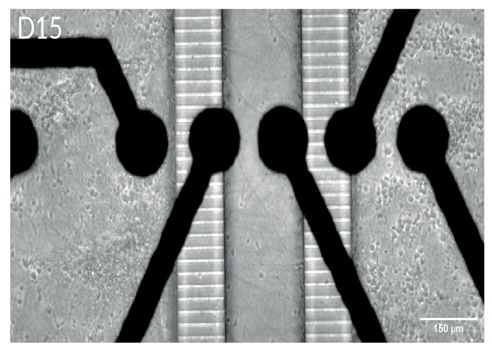
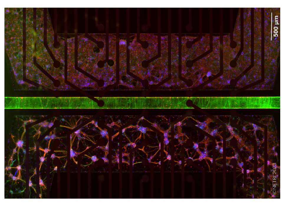
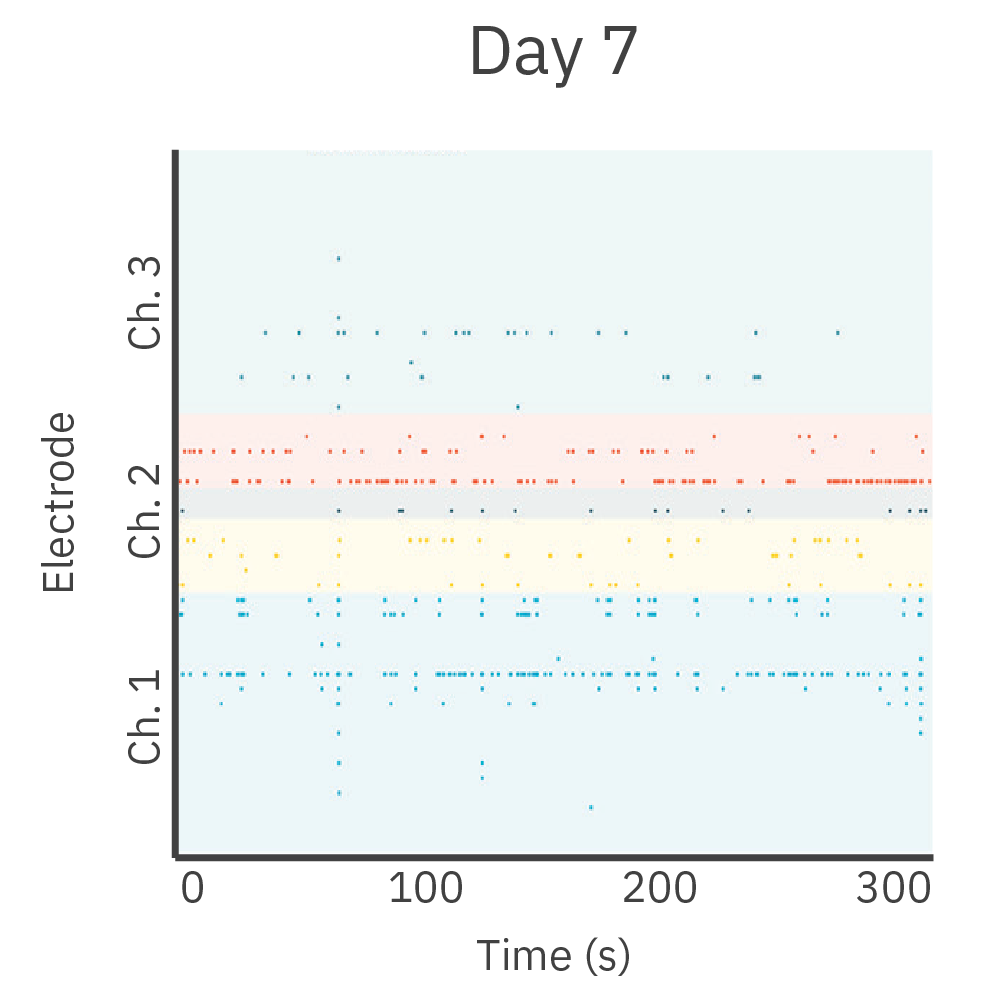
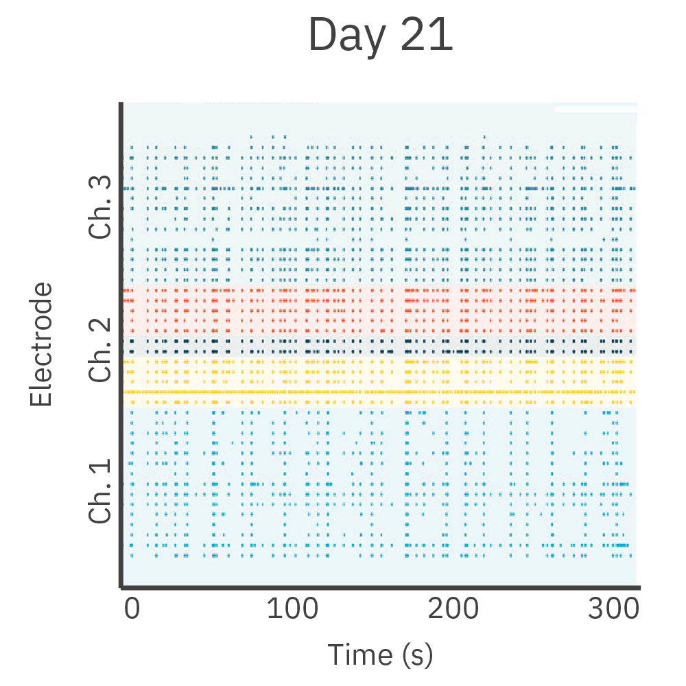
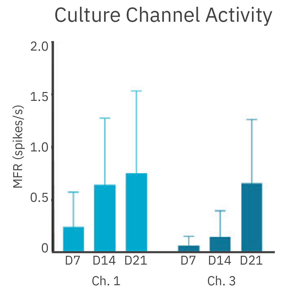
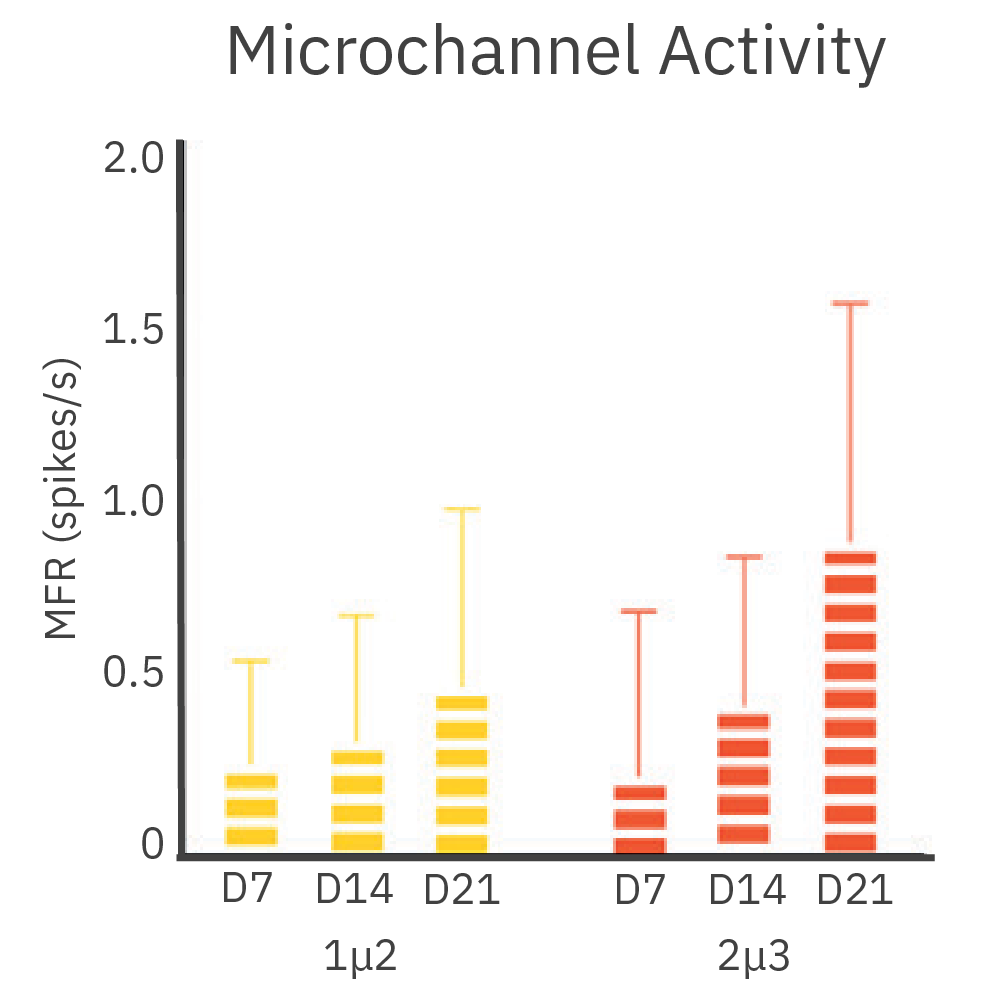
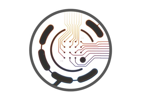
Cortical and hippocampal neurons were cultured in separate chambers of the DuaLink MEA (Netri) and recorded on the Maestro MEA platform over 21 days.
Results: Cultures increased synchronous firing between chambers as new synapses formed over time.
Purpose: To monitor growth and activity in neurites. Understanding the growth and function of neural projections could aid in the development neuroregenerative medicine.
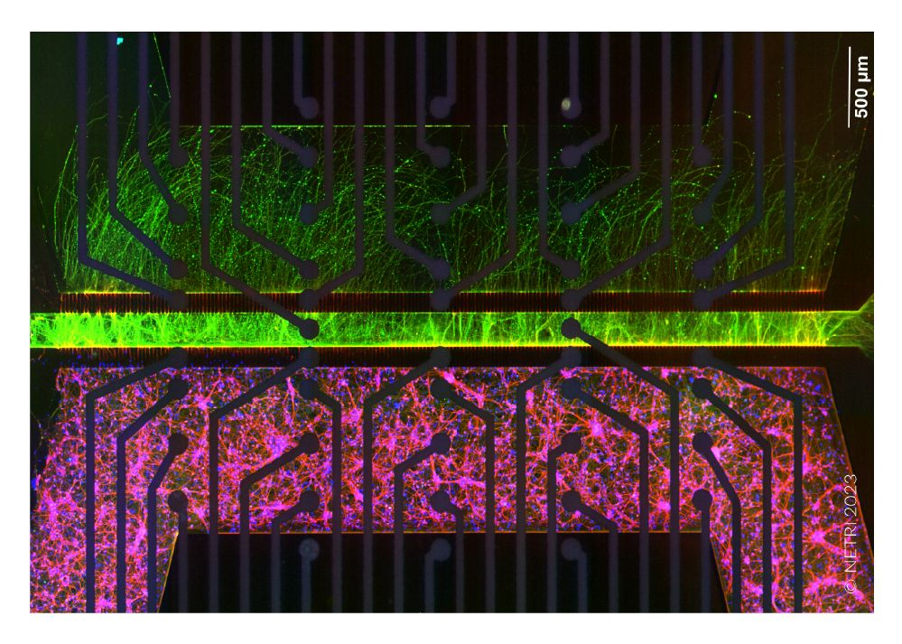
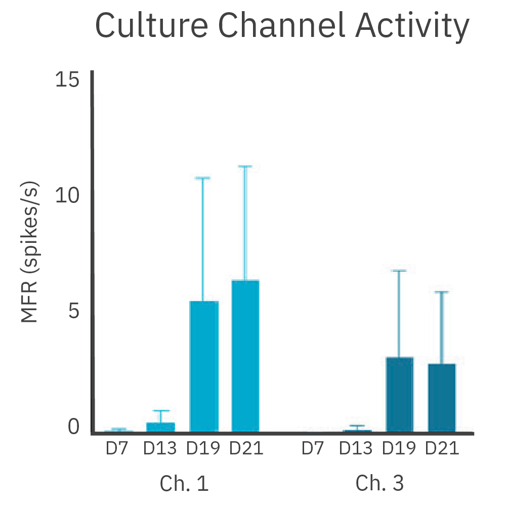
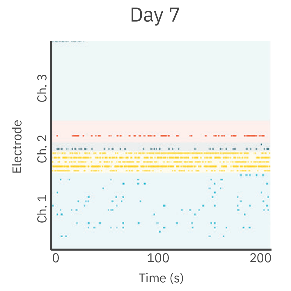
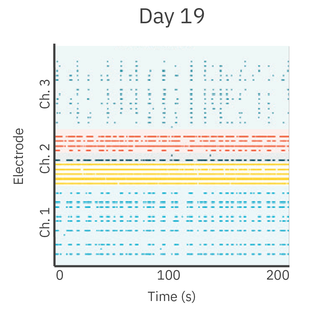
Cortical neurons were cultured in one chamber of the DuaLink MEA (Netri) and recorded on the Maestro MEA platform.
Results: As new neurite projections covered the empty chamber, increasing activity was observed. This activity was synchronous to the activity in the cortical chamber.
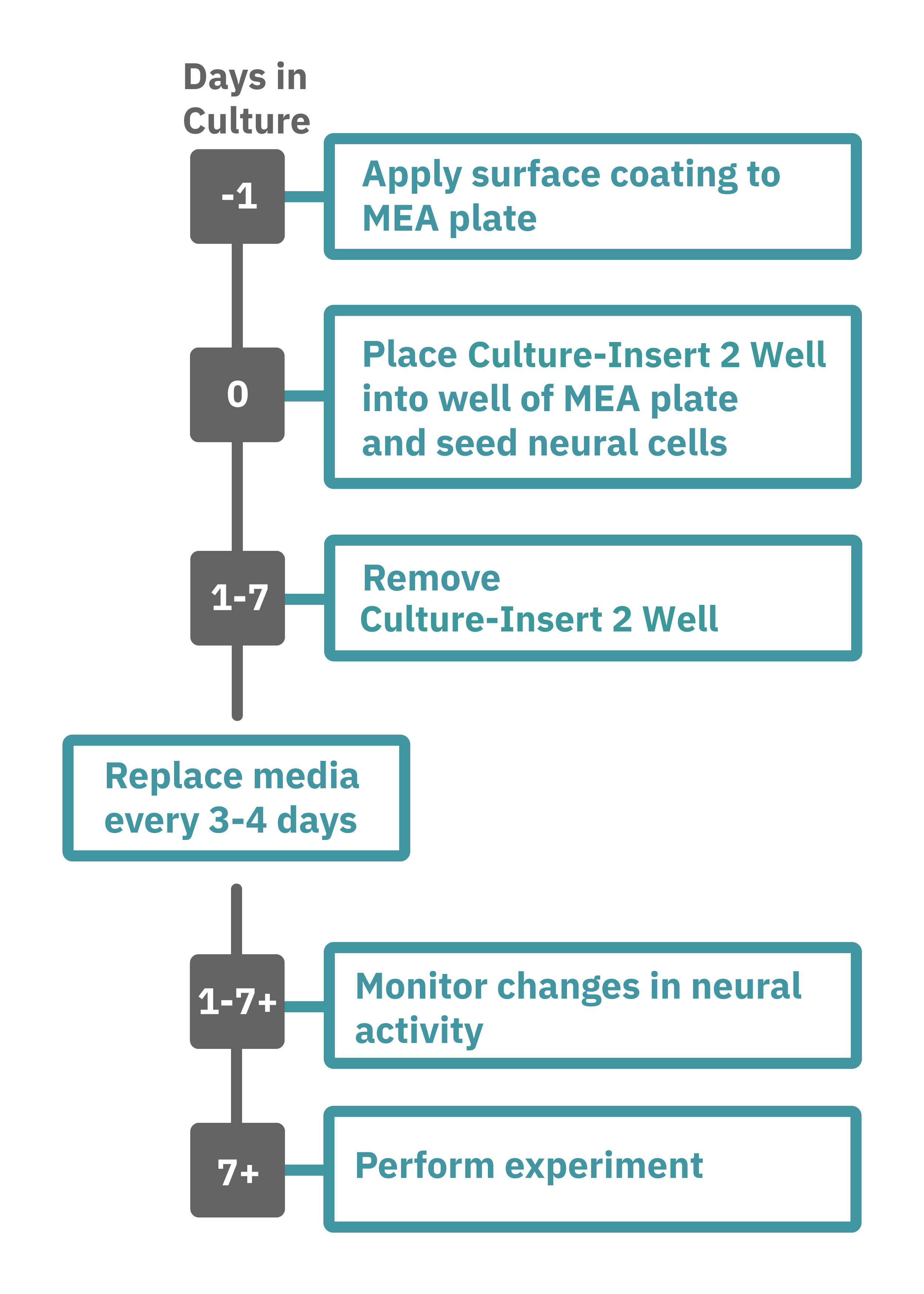
Getting started with Maestro Pro and Edge couldn't be easier. Place the Culture-Insert 2 Well (ibidi, 80209) into the well of a CytoView MEA 6 well plate (M384-tMEA-6W). Culture your neurons in the desired compartments of the Culture-Insert 2 Well (Day 0). Remove the Culture-Insert 2 Well after the cells have attached to the surface of the MEA plate (~1-7 days). Load the MEA plate into the Maestro MEA system and allow the environmental chamber to automatically equilibrate. Analyze the neural activity in the MEA plate label-free and in real-time with AxIS Navigator Neural Module software (Day 2+).